
 | ||
An exotoxin is a toxin secreted by bacteria. An exotoxin can cause damage to the host by destroying cells or disrupting normal cellular metabolism. They are highly potent and can cause major damage to the host. Exotoxins may be secreted, or, similar to endotoxins, may be released during lysis of the cell. Gram negative pathogens may secrete outer membrane vesicles containing lipopolysaccharide endotoxin and some virulence proteins in the bounding membrane along with some other toxins as intra-vesicular contents, thus adding a previously unforeseen dimension to the well-known eukaryote process of membrane vesicle trafficking, which is quite active at the host-pathogen interface.
Contents
- Types
- Type I cell surface active
- Superantigens
- Heat stable enterotoxins
- Type II membrane damaging
- Channel forming toxins
- Enzymatically active toxins
- Type III intracellular
- By mode of entry
- By mechanism
- Extracellular matrix damage
- References
They may exert their effect locally or produce systemic effects. Well-known exotoxins include: botulinum toxin produced by Clostridium botulinum; Corynebacterium diphtheriae toxin, produced during life-threatening symptoms of diphtheria; tetanospasmin produced by Clostridium tetani. The toxic properties of most exotoxins can be inactivated by heat or chemical treatment to produce a toxoid. These retain their antigenic specificity and can be used to produce antitoxins and, in the case of diphtheria and tetanus toxoids, are used as vaccines.
Exotoxins are susceptible to antibodies produced by the immune system, but many exotoxins are so toxic that they may be fatal to the host before the immune system has a chance to mount defenses against them. For this reason antitoxin, anti-serum containing antibodies, is injected to provide passive immunity.
Types
Many exotoxins have been categorized. This classification, while fairly exhaustive, is not the only system used. Other systems for classifying or identifying toxins include:
The same exotoxin may have different names, depending of the field of research.
Type I: cell surface-active
Type I toxins bind to a receptor on the cell surface and stimulate intracellular signaling pathways. Two examples are described below.
Superantigens
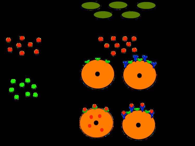
Superantigens are produced by several bacteria. The best-characterized superantigens are those produced by the strains of Staphylococcus aureus and Streptococcus pyogenes that cause toxic shock syndrome. Superantigens bridge the MHC class II protein on antigen-presenting cells with the T cell receptor on the surface of T cells with a particular Vβ chain. As a consequence, up to 50% of all T cells are activated, leading to massive secretion of proinflammatory cytokines, which produce the symptoms of toxic shock.
Heat-stable enterotoxins
Some strains of E. coli produce heat-stable enterotoxins (ST), which are small peptides that are able to withstand heat treatment at 100 °C. Different STs recognize distinct receptors on the cell surface and thereby affect different intracellular signaling pathways. For example, STa enterotoxins bind and activate membrane-bound guanylate cyclase, which leads to the intracellular accumulation of cyclic GMP and downstream effects on several signaling pathways. These events lead to the loss of electrolytes and water from intestinal cells.
Type II: membrane damaging
Membrane-damaging toxins exhibit hemolysin or cytolysin activity in vitro. However, induction of cell lysis may not be the primary function of the toxins during infection. At low concentrations of toxin, more subtle effects such as modulation of host cell signal transduction may be observed in the absence of cell lysis. Membrane-damaging toxins can be divided into two categories, the channel-forming toxins and toxins that function as enzymes that act on the membrane.
Channel-forming toxins
Most channel-forming toxins, which form pores in the target cell membrane, can be classified into two families: the cholesterol-dependent toxins and the RTX toxins.
Formation of pores by cholesterol-dependent cytolysins (CDC) requires the presence of cholesterol in the target cell. The size of the pores formed by members of this family is extremely large: 25-30 nm in diameter. All CDCs are secreted by the type II secretion system; the exception is pneumolysin, which is released from the cytoplasm of Streptococcus pneumoniae when the bacteria lyse.
The CDCs Streptococcus pneumoniae Pneumolysin, Clostridium perfringens perfringolysin O, and Listeria monocytogenes listeriolysin O cause specific modifications of histones in the host cell nucleus, resulting in down-regulation of several genes that encode proteins involved in the inflammatory response. Histone modification does not involve the pore-forming activity of the CDCs.
RTX toxins can be identified by the presence of a specific tandemly repeated nine-amino acid residue sequence in the protein. The prototype member of the RTX toxin family is haemolysin A (HlyA) of E. coli. RTX is also found in Legionella pneumophila.
Enzymatically active toxins
One example is the α toxin of C. perfringens, which causes gas gangrene; α toxin has phospholipase activity.
Type III: intracellular
Type III exotoxins can be classified by their mode of entry into the cell, or by their mechanism once inside.
By mode of entry
Intracellular toxins must be able to gain access to the cytoplasm of the target cell to exert their effects.
By mechanism
Once in the cell, many of the exotoxins act at the eukaryotic ribosomes (especially 60S), as protein synthesis inhibitors. (Ribosome structure is one of the most important differences between eukaryotes and prokaryotes, and, in a sense, these exotoxins are the bacterial equivalent of antibiotics such as clindamycin.)
Other intracellular toxins do not directly inhibit protein synthesis.
Extracellular matrix damage
These "toxins" allow the further spread of bacteria and, as a consequence, deeper tissue infections. Examples are hyaluronidase and collagenase. These molecules, however, are enzymes that are secreted by a variety of organisms and are not usually considered toxins. They are often referred to as virulence factors, since they allow the organisms to move deeper into the hosts tissues.