
 | ||
Shiga toxins are a family of related toxins with two major groups, Stx1 and Stx2, expressed by genes considered to be part of the genome of lambdoid prophages. The toxins are named for Kiyoshi Shiga, who first described the bacterial origin of dysentery caused by Shigella dysenteriae. The most common sources for Shiga toxin are the bacteria S. dysenteriae and the shigatoxigenic serotypes of Escherichia coli (STEC), which includes serotypes O157:H7, O104:H4, and other enterohemorrhagic E. coli (EHEC).
Contents
Nomenclature
Microbiologists use many terms to describe Shiga toxin and differentiate more than one unique form. Many of these terms are used interchangeably.
- Shiga toxin (Stx) – true Shiga toxin – is produced by Shigella dysenteriae.
- Shiga-like toxins 1 and 2 (SLT-1 and 2 or Stx-1 and 2) are the Shiga toxins produced by some E. coli strains. Stx-1 is identical to Stx or differs by only one amino acid. Stx-2 shares 56% sequence identity with Stx-1.
- Cytotoxins – an archaic denotation for Stx – is used in a broad sense.
- Verocytotoxins/verotoxins – a seldom-used term for Stx – is from the hypersensitivity of Vero cells to Stx.
Mechanism
Shiga toxins act to inhibit protein synthesis within target cells by a mechanism similar to that of ricin. After entering a cell via a macropinosome, the protein cleaves a specific adenine nucleobase from the 28S RNA of the 60S subunit of the ribosome, thereby halting protein synthesis.
Interestingly, the bacterial Shiga toxin can be used for targeted therapy of gastric cancer, because this tumor entity expresses the receptor of the Shiga toxin. For this purpose an unspecific chemotherapeutical is conjungated to the B-subunit to make it specific. In this way only the tumor cells, but not healthy cells should be destroyed during therapy.
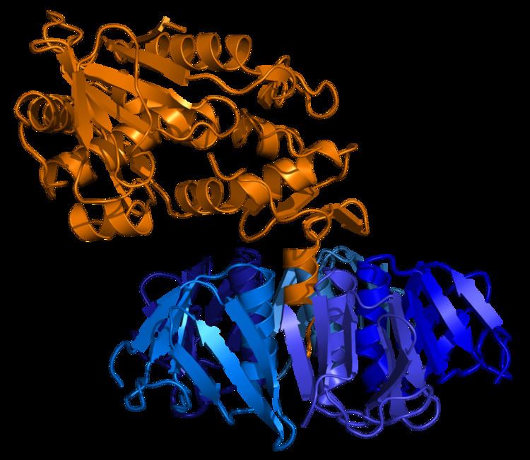
Structure
The toxin has two subunits—designated A (mol. wt. 32000 D) and B (mol. wt. 7700 D)—and is one of the AB5 toxins. The B subunit is a pentamer that binds to specific glycolipids on the host cell, specifically globotriaosylceramide (Gb3). Following this, the A subunit is internalised and cleaved into two parts. The A1 component then binds to the ribosome, disrupting protein synthesis. Stx-2 has been found to be about 400 times more toxic (as quantified by LD50 in mice) than Stx-1.
Gb3 is, for unknown reasons, present in greater amounts in renal epithelial tissues, to which the renal toxicity of Shiga toxin may be attributed. Gb3 is also found in central nervous system neurons and endothelium, which may lead to neurotoxicity. Stx-2 is also known to increase the expression of its receptor GB3 and cause neuronal dysfunctions.
The toxin requires highly specific receptors on the cells' surface to attach and enter the cell; species such as cattle, swine, and deer which do not carry these receptors may harbor toxigenic bacteria without any ill effect, shedding them in their feces, from where they may be spread to humans.