
ICD-10 L52 DiseasesDB 4462 eMedicine derm/138 | ICD-9-CM 695.2, 017.1 MedlinePlus 000881 | |
 | ||
Specialty | ||
Erythema nodosum (EN), also known as Subacute Migratory Panniculitis of Vilanova and Piñol, is an inflammatory condition characterised by inflammation of the fat cells under the skin, resulting in tender red nodules or lumps that are usually seen on both shins. It can be caused by a variety of conditions, and typically resolves spontaneously within 30 days. It is common in young people between 12–20 years of age.
Contents
Pre-eruptive phase
The first sign of erythema nodosum is often flu-like symptoms. It may involve a pyrexia, weakness and arthralgia.
Eruptive stage
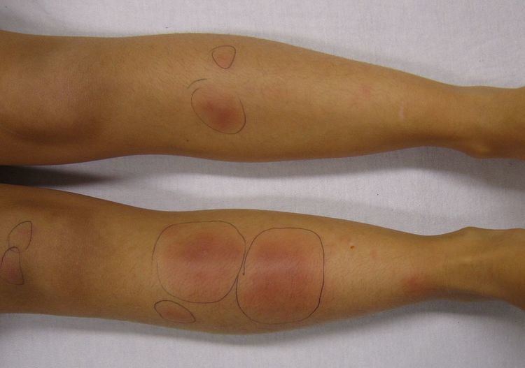
It is characterized by the appearance of dermo-hypodermic rounded nodules which may be bright red or purplish, bilaterally symmetrical, located on the extensor surface of the legs. They are movable relative to the deep plane and skin. They generally disappear in 1–6 weeks without sequelae.
Erythema nodosum is characterised by red nodules that are tender and warm. Nodules occur under the skin, classically in front of the shins. Nodules may occur anywhere there is fat under the skin, including the thighs, arms, trunk, face, and neck.
Erythema nodosum may occur concurrently with fever, malaise, and joint pain and inflammation. Nodules vary from 1–10 cm in diameter, and may coalesce to form large areas of hardened skin.
As the nodules age, they become bluish purple, brownish, yellowish, and finally green, similar to the color changes that occur in a resolving bruise. The nodules usually subside over a period of 2–6 weeks without ulceration or scarring.
Less common variants of erythema nodosum include:
Causes
EN is associated with a wide variety of conditions, including:
In about 30–50% of cases, the cause of EN is unknown.
EN may also be due to excessive antibody production in lepromatous leprosy leading to deposition of immune complexes.
There is an association with the HLA-B27 histocompatibility antigen, which is present in 65% of patients with erythema nodosum.
A useful mnemonic for causes is SORE SHINS (Streptococci, OCP, Rickettsia, Eponymous (Behçet), Sulfonamides, Hansen's Disease (Leprosy), IBD, NHL, Sarcoidosis.
Pathophysiology
Erythema nodosum is probably a delayed hypersensitivity reaction to a variety of antigens. Although circulating immune complexes have been demonstrated in patients with inflammatory bowel disease, they have not been found in idiopathic or uncomplicated cases.
Diagnosis
Erythema nodosum is diagnosed clinically. A biopsy can be taken and examined microscopically to confirm an uncertain diagnosis. Microscopic examination usually reveals a neutrophilic infiltrate surrounding capillaries that results in septal thickening, with fibrotic changes in the fat around blood vessels. A characteristic microscopic finding is radial granulomas, well-defined nodular aggregates of histiocytes surrounding a stellate cleft.
Additional evaluation should be performed to determine the underlying cause of erythema nodosum. This may include a full blood count, erythrocyte sedimentation rate (ESR), antistreptolysin-O (ASO) titer and throat culture, urinalysis, intradermal tuberculin test, and a chest x-ray. The ESR is typically high, the C-reactive protein elevated, and the blood showing an increase in white blood cells.
The ESR is initially very high, and falls as the nodules of erythema nodosum. The ASO titer is high in cases associated with a streptococcal throat infection. A chest X-ray should be performed to rule out pulmonary diseases, in particular sarcoidosis and Löfgren syndrome.
Treatment
Erythema nodosum is self-limiting and usually resolves itself within 3–6 weeks. A recurring form does exist, and in children it is attributed to repeated infections with streptococcus. Treatment should focus on the underlying cause. Symptoms can be treated with bedrest, leg elevation, compressive bandages, wet dressings, and nonsteroidal anti-inflammatory agents (NSAIDs). NSAIDs are usually more effective at the onset of EN versus with chronic disease.
Potassium iodide can be used for persistent lesions whose cause remains unknown. Corticosteroids and colchicine can be used in severe refractory cases. Thalidomide has been used successfully in the treatment of Erythema nodosum leprosum, and it was approved by the U.S. FDA for this use in July 1998.
Epidemiology
Erythema nodosum is the most common form of panniculitis. It is most common in the ages of 20–30, and affects women 3–6 times more than men.
About 15 percent of patients with inflammatory bowel disease develop erythema nodosum.
Eponym
The term, Subacute Migratory Panniculitis of Vilanova and Piñol, was named after the two famous Catalan Dermatologist who provided a brief description and explanation of the disease, Drs. Xavier Montiu Vilanova (1902–1965) and Joaquin Aguade Piñol (1918–1977), in 1954, and was named in 1956.