
Latin Epidermis TA A16.0.00.009 | Code TH H3.12.00.1.01001 FMA 70596 | |
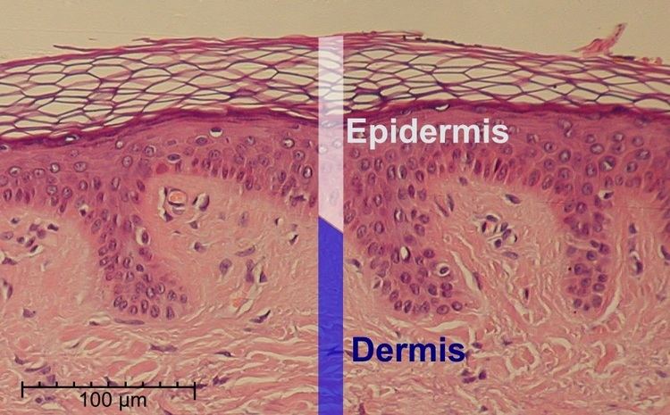 | ||
The epidermis is the outer (epi in Greek meaning "over" or "upon") of the two layers that make up the skin (or cutis), the inner layer being the dermis. It provides a barrier to infection from environmental pathogens and regulates the amount of water released from the body into the atmosphere through transepidermal water loss (TEWL). The outermost part of the epidermis is composed of stratified layers of flattened cells, that overlies a basal layer (stratum basale) composed of columnar cells arranged perpendicularly.
Contents
Cellular components
The epidermis has no blood supply and is nourished almost exclusively by diffused oxygen from the surrounding air. It is 95% keratinocytes (proliferating basal and differentiated suprabasal) but also contains melanocytes, Langerhans cells, Merkel cells, and inflammatory cells. Rete ridges ("rete tips") are epidermal thickenings that extend downward between dermal papillae. Blood capillaries are found beneath the epidermis, and are linked to an arteriole and a venule..
Layers
The epidermis is composed of 4 or 5 layers depending on the region of skin being considered. Those layers in descending order are:
The term Malpighian layer (stratum malpighi) is usually defined as both the stratum basale and stratum spinosum.
The epidermis is separated from the dermis, its underlying tissue, by a basement membrane.
Cell division
The stratified squamous epithelium is maintained by cell division within the stratum basale. Differentiating cell delaminate from the basement membrane and are displaced outwards through the epidermal layers, undergoing multiple stages of differentiation until, in the stratum corneum, losing their nucleus and fusing to squamous sheets, which are eventually shed from the surface (desquamation). Differentiated keratinocytes secrete keratin proteins which contribute to the formation of an extracellular matrix and is an integral part of the skin barrier function. In normal skin, the rate of keratinocyte production equals the rate of loss, taking about two weeks for a cell to journey from the stratum basale to the top of the stratum granulosum, and an additional four weeks to cross the stratum corneum. The entire epidermis is replaced by new cell growth over a period of about 48 days.
Calcium concentration
Keratinocyte differentiation throughout the epidermis is in part mediated by a calcium gradient, increasing from the stratum basale until the outer stratum granulosum, where it reaches its maximum, and decreasing in the stratum corneum. Calcium concentration in the stratum corneum is very low in part because those relatively dry cells are not able to dissolve the ions. This calcium gradient parallels keratinocyte differentiation and as such is considered a key regulator in the formation of the epidermal layers.
Elevation of extracellular calcium concentrations induces an increase in intracellular free calcium concentrations. Part of that intracellular increase comes from calcium released from intracellular stores and another part comes from transmembrane calcium influx, through both calcium-sensitive chloride channels and voltage-independent cation channels permeable to calcium. Moreover, it has been suggested that an extracellular calcium-sensing receptor (CaSR) also contributes to the rise in intracellular calcium concentration.
Development
Epidermal organogenesis, the formation of the epidermis, begins in the cells covering the embryo after neurulation, the formation of the central nervous system. In most vertebrates, this original one-layered structure quickly transforms into a two-layered tissue; a temporary outer layer, the periderm, which is disposed once the inner basal layer or stratum germinativum has formed.
This inner layer is a germinal epithelium that give rise to all epidermal cells. It divides to form the outer spinous layer (stratum spinosum). The cells of these two layers, together called the Malpighian layer(s) after Marcello Malpighi, divide to form the superficial granular layer (Stratum granulosum) of the epidermis.
The cells in the stratum granulosum do not divide, but instead form skin cells called keratinocytes from the granules of keratin. These skin cells finally become the cornified layer (stratum corneum), the outermost epidermal layer, where the cells become flattened sacks with their nuclei located at one end of the cell. After birth these outermost cells are replaced by new cells from the stratum granulosum and throughout life they are shed at a rate of 0.001 - 0.003 ounces of skin flakes every hour, or 0.024-0.072 ounces per day.
Epidermal development is a product of several growth factors, two of which are:
Barrier
The epidermis serves as a barrier to protect the body against microbial pathogens, oxidant stress (UV light) and chemical compounds and provides mechanical resistance. Most of that function is played by the stratum corneum.
Characteristics of the barrier
Factors that alter the barrier
Skin hydration
The ability of the skin to hold water is primarily due to the stratum corneum and is critical for maintaining healthy skin. Lipids arranged through a gradient and in an organized manner between the cells of the stratum corneum form a barrier to transepidermal water loss.
Skin color
The amount and distribution of melanin pigment in the epidermis is the main reason for variation in skin color in Homo sapiens. Melanin is found in the small melanosomes, particles formed in melanocytes from where they are transferred to the surrounding keratinocytes. The size, number, and arrangement of the melanosomes varies between racial groups, but while the number of melanocytes can vary between different body regions, their numbers remain the same in individual body regions in all human beings. In white and Asian skin the melanosomes are packed in "aggregates", but in black skin they are larger and distributed more evenly. The number of melanosomes in the keratinocytes increases with UV radiation exposure, while their distribution remain largely unaffected.
Clinical significance
Laboratory culture of keratinocytes to form a 3D structure (artificial skin) recapitulating most of the properties of the epidermis is routinely used as a tool for drug development and testing.