
Latin venula TA A12.0.00.037 | Code TH H3.09.02.0.03002 FMA 63130 | |
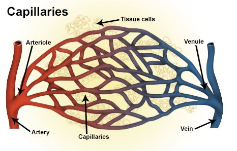 | ||
A venule is a very small blood vessel in the microcirculation that allows blood to return from the capillary beds to drain into the larger blood vessels, the veins. Venules range from 7 to 50μm in diameter. Veins contain approximately 70% of total blood volume, 25% of which is contained in the venules. Many venules unite to form a vein.
Structure
Venule walls have three layers: An inner endothelium composed of squamous endothelial cells that act as a membrane, a middle layer of muscle and elastic tissue and an outer layer of fibrous connective tissue. The middle layer is poorly developed so that venules have thinner walls than arterioles. They are extremely porous so that fluid and blood cells can move easily from the bloodstream through their walls.
In contrast to regular venules, high endothelial venules are a special type of venule where the endothelium is made up of simple cuboidal cells. Lymphocytes exit the blood stream and enter the lymph nodes via these specialized venules when an infection is detected.
They form from anastomosis of capillaries, using the beta form of semi-red blood cells and white blood cells to form.