
DiseasesDB 32954 | ||
 | ||
A dural arteriovenous fistula (DAVF), is an abnormal direct connection (fistula) between a meningeal artery and a meningeal vein or dural venous sinus. In cases where there are multiple fistulas, the related term dural arteriovenous malformation (DAVF) is used.
Contents
Signs and Symptoms
The most common signs/symptoms of DAVFs are:
- Pulsatile tinnitus
- Occipital bruit
- Headache
- Visual impairment
- Papilledema
Pulsatile tinnitus is the most common symptom in patients, and it is associated with transverse-sigmoid sinus DAVFs. Carotid-cavernous DAVFs, on the other hand, are more closely associated with pulsatile exophthalmos. DAVFs may also be asymptomatic (e.g. cavernous sinus DAVFs).
Location
Most commonly found adjacent to dural sinuses in the following locations:
- Transverse (lateral) sinus, left-sided slightly more common than right
- Intratentorial
- From the posterior cavernous sinus, usually draining to the transverse or sigmoid sinuses
- Vertebral artery (posterior meningeal branch)
Causes
It is still unclear whether DAVFs are congenital or acquired. Current evidence supports transverse-sigmoid sinus junction dural malformations are acquired defects, occurring in response to thrombosis and collateral revascularization of a venous sinus.
Diagnosis
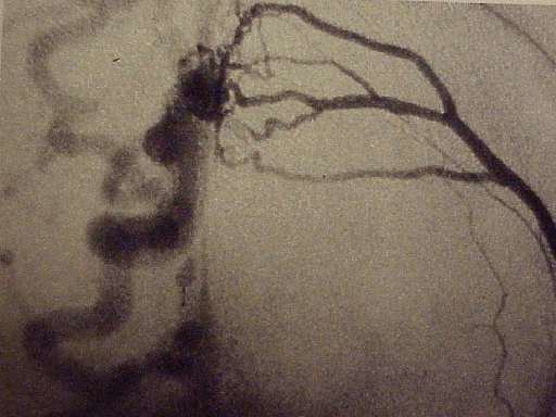
Cerebral angiography is the diagnostic standard. MRIs are usually normal.
Borden Classification
The Borden Classification of dural arteriovenous malformations or fistulas, groups into three types based upon their venous drainage:
- Type I: dural arterial supply drains anterograde into venous sinus.
- Type II: dural arterial supply drains into venous sinus. High pressure in sinus results in both anterograde drainage and retrograde drainage via subarachnoid veins.
- Type III: dural arterial supply drains retrograde into subarachnoid veins.
Type I
Type I dural arteriovenous fistulas are supplied by meningeal arteries and drain into a meningeal vein or dural venous sinus. The flow within the draining vein or venous sinus is anterograde.
- Type Ia – simple dural arteriovenous fistulas have a single meningeal arterial supply
- Type Ib – more complex arteriovenous fistulas are supplied by multiple meningeal arteries
The distinction between Types Ia and Ib is somewhat specious as there is a rich system of meningeal arterial collaterals. Type I dural fistulas are often asymptomatic, do not have a high risk of bleeding and do not necessarily need to be treated.
Type II
The high pressure within a Type II dural AV fistula causes blood to flow in a retrograde fashion into subarachnoid veins which normally drain into the sinus. Typically this is because the sinus has outflow obstruction. Such draining veins form venous varices or aneurysms which can bleed. Type II fistulas need to be treated to prevent hemorrhage. The treatment may involve embolization of the draining sinus as well as clipping or embolization of the draining veins.
Type III
Type III dural AV fistulas drain directly into subarachnoid veins. These veins can form aneurysms and bleed. Type III dural fistulas need to be treated to prevent hemorrhage. Treatment can be as simple as clipping the draining vein at the site of the dural sinus. If treatment involves embolization, it will only typically be effective if the glue traverses the actual fistula and enters, at least slightly, the draining vein.
Cognard et al. Classification
The Cognard et al. Classification correlates venous drainage patterns with increasingly aggressive neurological clinical course.
Indications
Embolization
One approach used for treatment is embolization. A six-vessel angiogram is employed to determine the vascular supply to the fistula. Detachable coils, liquid embolic agents like NBCA, and onyx, or combinations of both are injected into the blood vessel to occlude the DAVF. Preoperative embolization can also be used to supplement surgery.
Surgery
DAVFs are also managed surgically. The operative approach varies depending on the location of the lesion.
Stereotactic radiosurgery
Stereotactic radiosurgery is used obliterating DAVFs post-embolization, and is considered an important adjunct. Use of this method, however, is limited to benign DAVFs that have failed other treatments.
Epidemiology
10-15% of intracranial AV malformations are DAVFs. There is a higher preponderance in females (61-66%), and typically patients are in their fourth or fifth generation of life. DAVFs are rarer in children.
Research Directions
Manual carotid self compression is a controversial treatment for DAVF. Patients using this method are told to compress the carotid with the opposite hand for approximately 10 minutes daily, and gradually increasing the frequency and duration of compression. Currently, it is unclear whether this method is an effective therapy.