
Specialty Neurosurgery ICD-9-CM 377.0 eMedicine oph/187 | ICD-10 H47.1 DiseasesDB 9580 Patient UK Papilledema | |
 | ||
Papilledema (or papilloedema) is optic disc swelling that is caused by increased intracranial pressure. The swelling is usually bilateral and can occur over a period of hours to weeks. Unilateral presentation is extremely rare. Papilledema is mostly seen as a symptom resulting from another pathophysiological process.
Contents
In intracranial hypertension, papilledema most commonly occurs bilaterally. When papilledema is found on fundoscopy, further evaluation is warranted as vision loss can result if the underlying condition is not treated. Further evaluation with a CT or MRI of the brain and/or spine is usually performed. Recent research has shown that point-of-care ultrasound can be used to measure optic nerve sheath diameter for detection of increased intracranial pressure and shows good diagnostic test accuracy compared to CT. Thus, if there is a question of papilledema on fundoscopic examination or if the optic disc cannot be adequately visualized, ultrasound can be used to rapidly assess for increased intracranial pressure and help direct further evaluation and intervention. Unilateral papilledema can suggest a disease in the eye itself, such as an optic nerve glioma.
Signs and symptoms
Papilledema may be asymptomatic or present with headache in the early stages. However it may progress to enlargement of the blind spot, blurring of vision, visual obscurations (inability to see in a particular part of the visual field for a period of time) and ultimately total loss of vision may occur.
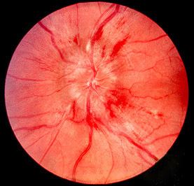
The signs of papilledema that are seen using an ophthalmoscope include:
On visual field examination, the physician may elicit an enlarged blind spot; the visual acuity may remain relatively intact until papilledema is severe or prolonged.
Diagnosis
Checking the eyes for signs of papilledema should be carried out whenever there is a clinical suspicion of raised intracranial pressure, and is recommended in newly onset headaches. This may be done by ophthalmoscopy or fundus photography, and possibly slit lamp examination.
Causes
Raised intracranial pressure as a result of one or more of the following:
Pathophysiology
As the optic nerve sheath is continuous with the subarachnoid space of the brain (and is regarded as an extension of the central nervous system), increased pressure is transmitted through to the optic nerve. The brain itself is relatively spared from pathological consequences of high pressure. However, the anterior end of the optic nerve stops abruptly at the eye. Hence the pressure is asymmetrical and this causes a pinching and protrusion of the optic nerve at its head. The fibers of the retinal ganglion cells of the optic disc become engorged and bulge anteriorly. Persistent and extensive optic nerve head swelling, or optic disc edema, can lead to loss of these fibers and permanent visual impairment.
Treatment
Historically, papilledema was a potential contraindication to lumbar puncture, as it indicates a risk for tentorial herniation and subsequent death via cerebral herniation, however newer imaging techniques have been more useful at determining when and when not to conduct a lumbar puncture. Imaging by CT or MRI is usually performed to elicit whether there is a structural cause i.e., tumor. An MRA and MRV may also be ordered to rule out the possibility of stenosis or thrombosis of the arterial or venous systems.
The treatment depends largely on the underlying cause. However, the root cause of papilledema is the increased intracranial pressure (ICP). This is a dangerous sign, indicative of a brain tumor, CNS inflammation or idiopathic intracranial hypertension (IIH) that may become manifest in the near future.
Thus, a biopsy is routinely performed prior to the treatment in the initial stages of papilledema to detect whether a brain tumor is present. If detected, laser treatment, radiation and surgeries can be used to treat the tumor.
To decrease ICP, medications can be administered by increasing the absorption of Cerebrospinal fluid (CSF), or decreasing its production. Such medicines include diuretics like acetazolamide and furosemide. These diuretics, along with surgical interventions, can also treat IIH. In IIH, weight loss (even a loss of 10-15%) can lead to normalization of ICP.
Meanwhile, steroids can reduce inflammation (if this is a contributing factor to increased ICP), and may help to prevent vision loss. However, steroids have also been known to cause increased ICP, especially with a change in dosage. However, if a severe inflammatory condition exists, such as multiple sclerosis, steroids with anti-inflammatory effects such as Methylprednisolone and prednisone can help.
Other treatments include repeated lumbar punctures to remove excess spinal fluid in the cranium. The removal of potentially causative medicines including tetracyclines and vitamin A analogues may help decrease ICP; however, this is only necessary if the medication is truly felt to contribute to the ICP increase.