
Latin Sinus durae matris Dorlands/Elsevier s_12/12738708 FMA 76590 | MeSH A07.231.908.224 TA A12.3.05.101 | |
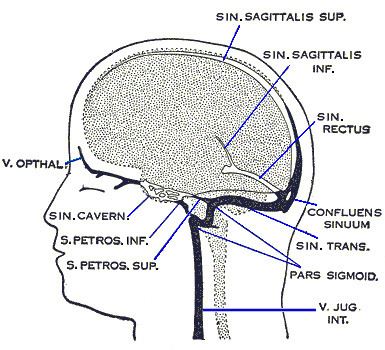 | ||
The dural venous sinuses (also called dural sinuses, cerebral sinuses, or cranial sinuses) are venous channels found between the periosteal and meningeal layers of dura mater in the brain. They receive blood from internal and external veins of the brain, receive cerebrospinal fluid (CSF) from the subarachnoid space via arachnoid granulations, and mainly empty into the internal jugular vein.
Contents
Structure
The walls of the dural venous sinuses are composed of dura mater lined with endothelium, a specialized layer of flattened cells found in blood vessels. They differ from other blood vessels in that they lack a full set of vessel layers (e.g. tunica media) characteristic of arteries and veins. It also lacks valves as seen in veins.
Clinical relevance
The sinuses can be injured by trauma in which damage to the dura mater, may result in blood clot formation (thrombosis) within the dural sinuses. Other common causes of dural sinus thrombosis include tracking of infection through the ophthalmic vein in orbital cellulitis. While rare, dural sinus thrombosis may lead to hemorrhagic infarction or cerebral oedema with serious consequences including epilepsy, neurological deficits, or death.