
 | ||
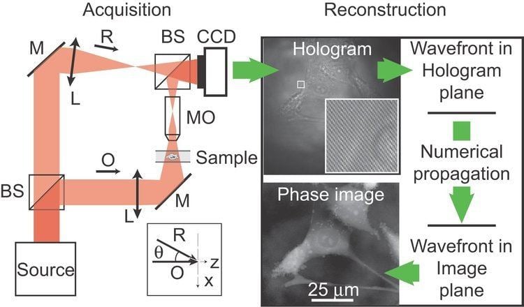
Digital Holographic Microscopy (DHM) is digital holography applied to microscopy. Digital holographic microscopy distinguishes itself from other microscopy methods by not recording the projected image of the object. Instead, the light wave front information originating from the object is digitally recorded as a hologram, from which a computer calculates the object image by using a numerical reconstruction algorithm. The image forming lens in traditional microscopy is thus replaced by a computer algorithm. Other closely related microscopy methods to digital holographic microscopy are interferometric microscopy, optical coherence tomography and diffraction phase microscopy. Common to all methods is the use of a reference wave front to obtain amplitude (intensity) and phase information. The information is recorded on a digital image sensor or by a photo detector from which an image of the object is created (reconstructed) by a computer. In traditional microscopy, which do not use a reference wave front, only intensity information is recorded and essential information about the object is lost.
Contents
- How interactive visualization led to insights in digital holographic microscopy scipy 2014 rebec
- Working principle
- Advantages
- Applications
- Living Cells Imaging by DHM
- Surface 3D topography
- Time resolved applications
- MEMS
- Metrology
- Industrial inspection
- Micro optics
- 3D particle tracking
- History
- References
Holography was invented by Dennis Gabor to improve electron microscopy. Nevertheless, it never found many concrete and industrial applications in this field.
Actually, DHM has been mostly applied to light microscopy. It has shown in this field unique applications for 3D characterization of technical samples and enables quantitative characterization of living cells. In material Science, DHM are used routinely for research in academic and industrial labs, depending of the application microscopes can be configured both in transmission and in reflection. DHM is a unique solution for 4D (3D + time) characterization of technical samples, when information needs to be grabbed very quickly. It is the case for measurements in noisy environment, in presence of vibrations, when the samples move, or when the shape of samples change due to external stimuli, such as mechanical, electrical, or magnetic forces, chemical erosion or deposition, evaporation, … . In life sciences, DHM are usually configured in transmission mode. They enable label free Quantitative Phase Measurement (QPM), also called Quantitative Phase Imaging (QPI), of living cells. Measurements do not affect the cells, enabling long term studies. It provides information that can be interpreted into many underlying biological processes as explained in the section “Living Cells Imaging” here below.
How interactive visualization led to insights in digital holographic microscopy scipy 2014 rebec
Working principle
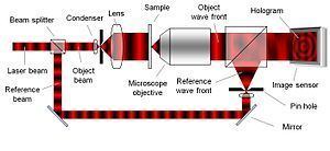
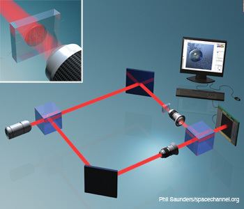
To create the necessary interference pattern, i.e., the hologram, the illumination needs to be a coherent (monochromatic) light source, a laser for example. As can be seen in Figure 2, the laser light is split into an object beam and a reference beam. The expanded object beam illuminates the sample to create the object wave front. After the object wave front is collected by a microscope objective, the object and reference wave fronts are joined by a beam splitter to interfere and create the hologram. Using the digitally recorded hologram, a computer acts as a digital lens and calculates a viewable image of the object wave front by using a numerical reconstruction algorithm.
Commonly, a microscope objective is used to collect the object wave front. However, as the microscope objective is only used to collect light and not to form an image, it may be replaced by a simple lens. If a slightly lower optical resolution is acceptable, the microscope objective may be entirely removed.
Digital holography comes in different flavors, such as off-axis Fresnel, Fourier, image plane, in-line, Gabor and phase-shifting digital holography, depending on the optical setup. The basic principle, however, is the same; a hologram is recorded and an image is reconstructed by a computer.
The lateral optical resolution of digital holographic microscopy is equivalent to the resolution of traditional light microscopy. DHM is diffraction-limited by the numerical aperture, in the same way as traditional light microscopy. However, DHM offers a superb axial (depth) resolution. An axial accuracy of approximately 5 nm has been reported.
Advantages
Phase shift images
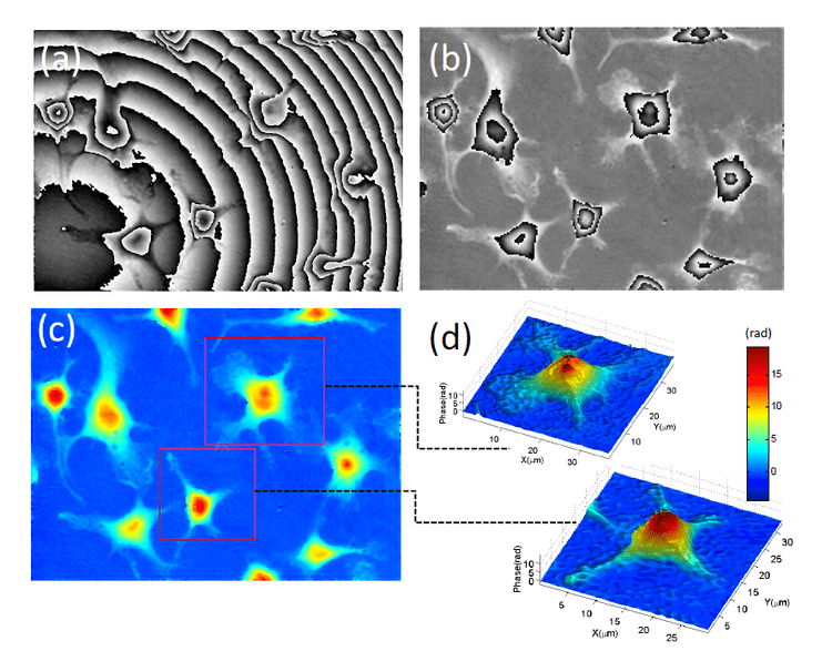
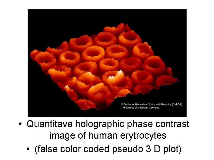
Besides the ordinary bright field image, a phase shift image is created as well. The phase shift image is unique for digital holographic microscopy and gives quantifiable information about optical distance. In reflection DHM, the phase shift image forms a topography image of the object.
Transparent objects, like living biological cells, are traditionally viewed in a phase contrast microscope or in a differential interference contrast microscope. These methods visualize phase shifting transparent objects by distorting the bright field image with phase shift information. Instead of distorting the bright field image, transmission DHM creates a separate phase shift image showing the optical thickness of the object. Digital holographic microscopy thus makes it possible to visualize and quantify transparent objects and is therefore also referred to as quantitative phase contrast microscopy.
Traditional phase contrast or bright field images of living unstained biological cells, Figure 3 (right), have proved themselves to be very difficult to analyze with image analysis software. On the contrary, phase shift images, Figure 3 (left), are readily segmented and analyzed by image analysis software based on mathematical morphology, such as CellProfiler.
3-Dimensional information
An object image is calculated at a given focal distance. However, as the recorded hologram contains all the necessary object wave front information, it is possible to calculate the object at any focal plane by changing the focal distance parameter in the reconstruction algorithm. In fact, the hologram contains all the information needed to calculate a complete image stack. In a DHM system, where the object wave front is recorded from multiple angles, it is possible to fully characterize the optical characteristics of the object and create tomography images of the object.
Digital autofocus
Conventional autofocus is achieved by vertically changing the focal distance until a focused image plane is found. As the complete stack of image planes may be calculated from a single hologram, it is possible to use any passive autofocus method to digitally select the focal plane. The digital auto focusing capabilities of digital holography opens up the possibility to scan and image surfaces extremely rapidly, without any vertical mechanical movement. By recording a single hologram and afterwards stitch sub-images together that are calculated at different focal planes, a complete and focused image of the object may be created.
Optical aberration correction
As DHM systems do not have an image forming lens, traditional optical aberrations do not apply to DHM. Optical aberrations are "corrected" by design of the reconstruction algorithm. A reconstruction algorithm that truly models the optical setup will not suffer from optical aberrations.
Low cost
In optical microscopy systems, optical aberrations are traditionally corrected by combining lenses into a complex and costly image forming microscope objective. Furthermore, the narrow focal depth at high magnifications requires precision mechanics. The needed components for a DHM system are inexpensive optics and semiconductor components, such as a laser diode and an image sensor. The low component cost in combination with the auto focusing capabilities of DHM, make it possible to manufacture DHM systems for a very low cost.
Applications
Digital holographic microscopy has been successfully applied in a range of application areas.
Living Cells Imaging by DHM
However, due to DHM’s capability of non-invasively visualizing and quantifying biological tissue, bio-medical applications have received most attention. Examples of bio-medical applications are:
Surface 3D topography
DHM performs static measurements of 3D surface topography as many other 3D optical profilometers (white light interferometers, confocal, focus variation, … ). It enables to retrieve, roughness and shape of many surfaces. Use of multiple wavelengths enable to overcome the l/4 limit of traditional phase shifting interferometers. Applications have been demonstrated on many samples such as medical implants, watch components, micro components, micro-optics.
Time resolved applications
As DHM measures the 3D surface topography over the full field of view within a single camera acquisition. There is no need for scanning, neither vertical, nor lateral. Consequently, dynamic changes of topography are measured instantaneously. The acquisition rate is only limited by the camera frame. Measurement have demonstrated on many type of samples such as smart surface, self-healing surfaces, not equilibrium systems, evaporation processes, electrodeposition, evaporation, crystallization, mechanical deformation, …
MEMS
Use in conjunction with a stroboscopic electronic unit to synchronize the laser pulse for sample illumination and the camera acquisition with the MEMS excitation, DHM® provides time sequences of 3D topography along the excitation phase of the microsystems. Analysis of this time sequence of 3D topographies acquired at a fixed frequency provides vibration map and enable decomposition of the movement in term of in- and out-of-plane.
Sweeping of the excitation frequency provides structural resonances as well as amplitude and phase Bode analysis. Measurement have demonstrated on many type of MEMS such as comb drive actuators, micro-mirrors, accelerometers, gyroscopes, micro pumps, microphones, ultrasonic transducers, cantilevers, and surface acoustic waves among others.
Metrology
DHM refers only to wavelengths for height measurement. Therefore, DHM provides precise height measurements with very high repeatability and linearity independently of any vertical calibration, precise positioning of mechanical part, repeatability of interferometric piezo-controller, motorized displacement, or liquid crystal display scanning. This feature makes out of DHM an outstanding tool for step and roughness certification among other. For transmission systems, perfect flatness calibration is achieved by taking as reference an acquisition without any sample in the optical path. Flatness calibration of reflection type systems requires the use of a perfectly flat sample.
Industrial inspection
The very short time needed to grab information makes DHM very robust to environmental vibrations. It enables in particular “on-flight” and “on-line” quality controls of parts. Applications have been demonstrated in particular for implants roughness, structure of semiconductor components, solar industry, industrial metrology, and watch parts among other.
Micro optics
Micro optics arrays fast measurement and inspection have been demonstrated and compared successfully with measurement made with other profilometers.
Extended depth of focus algorithms based on digital focalization enables have a sharp focus over the full lens surface, even for high NA samples. DHM has been also applied to dynamical characterization of variable lenses.
3D particle tracking
3D particle tracking has been demonstrated in numerous publications [to be completed]. A Z-stack of measurement can be reconstructed digitally from a single hologram using a range of propagation distances. Specific algorithms enable to determine for each particle the distance corresponding to its best focus. Performing this operation on a time-sequence of holograms enables to determine the trajectories of particles.
History
The first reports of replacing the photographic hologram of classical holography by digitally recording the hologram and numerically reconstructing the image in a computer was published in the late 1960s and in the early 1970s. Similar ideas were proposed for the electron microscope in the early 1980s. But, computers were too slow and recording capabilities were too poor for digital holography to be useful in practice. After the initial excitement, digital holography went into a similar hibernation as holography experienced about two decades earlier.
In the mid 1990s, digital image sensors and computers had become powerful enough to reconstruct images with some quality. In the 1960s, digital holography could either mean to compute an image from a hologram or to compute a hologram from a 3D model. The latter developed in parallel with classical holography during the hibernation of digital holography. During that time, digital holography was synonymous with what is now known as computer generated holography.
By the mid 1990s, digital image sensors and computers had improved tremendously, but still lacked the required pixel count and density for digital holography to be anything more than a curiosity. At the time, the market driving digital image sensors was primarily low-resolution video, and so those sensors provided only PAL, NTSC, or SECAM resolution. This suddenly changed at the beginning of the 21st century with the introduction of digital still image cameras, which drove demand for inexpensive high-pixel-count sensors. As of 2010, affordable image sensors can have up to 60 megapixels. In addition, the CD and DVD-player market has driven development of affordable diode lasers and optics.
The first reports of using digital holography for light microscopy came in the mid 1990s. However, it was not until the early 2000s that image sensor technology had progressed far enough to allow images of a reasonable quality. During this time, the first commercial digital holographic microscopy companies, 4Deep inwater imaging, Lyncée tec, and Phase Holographic Imaging, were founded. With increased computing power and use of inexpensive high-resolution sensors and lasers, digital holographic microscopy is today finding applications primarily within life science, oceanology and metrology.