
 | ||
The haemocytometer is a device usually used to count blood cells. It was originally designed for the counting of blood cells.
Contents
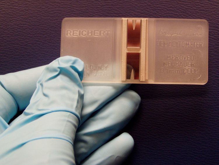
The haemocytometer was invented by Louis-Charles Malassez and consists of a thick glass microscope slide with a rectangular indentation that creates a chamber. This chamber is engraved with a laser-etched grid of perpendicular lines. The device is carefully crafted so that the area bounded by the lines is known, and the depth of the chamber is also known. It is therefore possible to count the number of cells or particles in a specific volume of fluid, and thereby calculate the concentration of cells in the fluid overall.
Principles
The gridded area of the haemocytometer consists of nine 1 x 1 mm (1 mm2) squares. These are subdivided in 3 directions; 0.25 x 0.25 mm (0.0625 mm2), 0.25 x 0.20 mm (0.05 mm2) and 0.20 x 0.20 mm (0.04 mm2). The central square is further subdivided into 0.05 x 0.05 mm (0.0025 mm2) squares. The raised edges of the hemocytometer hold the coverslip 0.1 mm off the marked grid, giving each square a defined volume (see figure on the right).
Usage
To use the hemocytometer, first make sure that the special coverslip provided with the counting chamber is properly positioned on the surface of the counting chamber. When the two glass surfaces are in proper contact Newton's rings can be observed. If so, the cell suspension is applied to the edge of the coverslip to be sucked into the void by capillary action which completely fills the chamber with the sample. The number of cells in the chamber can be determined by direct counting using a microscope, and visually distinguishable cells can be differentially counted. The number of cells in the chamber is used to calculate the concentration or density of the cells in the mixture the sample comes from. It is the number of cells in the chamber divided by the chamber's volume, which is known from the start, taking account of any dilutions and counting shortcuts:
where the volume of the diluted sample (after dilution) divided by the volume of the original mixture in the sample (before dilution) is the dilution factor. For example, if the volume of the original mixture was 20μL and it was diluted once (by adding 20μL dilutant), then the second term in parentheses is 40μL/20μL. The volume of the squares counted is the one shown in the table at the top, depending on the size (see figure on the right). The number of cells counted is the sum of all cells counted across squares in one chamber. The proportion of the cells counted applies if not all inner squares within a set square are counted (i.e., if only 4 out of the 20 in a corner square are counted, then this term will equal 0.2).
For most applications, the four large corner squares are only used. The cells that are on or touching the top and left lines are counted, but the ones on or touching the right or bottom lines are ignored.