
Order Unassigned Rank Species | Higher classification Dengue virus group | |
 | ||
Similar Aedes, Yellow fever mosquito, Asian tiger mosquito, Flaviviridae, Mosquito | ||
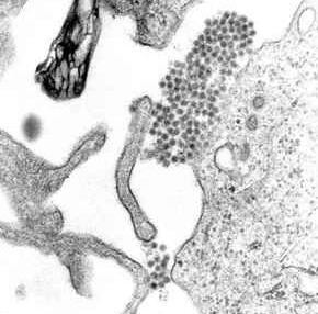
Dengue virus (DENV) is the cause of dengue fever. It is a mosquito-borne single positive-stranded RNA virus of the family Flaviviridae; genus Flavivirus. Five serotypes of the virus have been found, all of which can cause the full spectrum of disease. Nevertheless, scientists are finding their understanding of dengue virus may be simplistic, as rather than distinct antigenic groups there appears to be a continuum. This same study identified 47 strains of dengue virus. Additionally, coinfection with and lack of rapid tests for zika virus and chikungunya complicate matters in real world infections.
Contents
- Dengue virus life cycle youtube vti swl 04
- Evolution
- Life cycle
- E protein
- prMM protein
- NS3 protein
- NS5 protein
- Complexes between the E protein and neutralizing antibodies
- Mechanism of Infection
- Severe disease
- Immune system interaction
- Inhibition of interferon signaling by blocking signal transducer
- Inhibition of the type I interferon response
- Aedes aegypti D7 Saliva Protein
- Vaccine research
- References
Its genome is about 11000 bases of positive-sense single stranded RNA (ssRNA) that codes for three structural proteins (capsid protein C, membrane protein M, envelope protein E) and seven nonstructural proteins (NS1, NS2a, NS2b, NS3, NS4a, NS4b, NS5). It also includes short non-coding regions on both the 5' and 3' ends.
Dengue virus life cycle youtube vti swl 04
Evolution
The dengue type 1 virus appears to have evolved in the early 19th century. Based on the analysis of the envelope protein there are at least four genotypes (1 to 4). In 2013 a fifth serotype was reported. The rate of nucleotide substitution for this virus has been estimated to be 6.5×10⁻⁴ per nucleotide per year, a rate similar to other RNA viruses. The American African genotype has been estimated to have evolved between 1907 and 1949. This period includes World Wars I and II, which were associated with considerable movement of populations and environmental disturbance, factors known to promote the evolution of new vector-borne viral species.
Life cycle
Until a few hundred years ago dengue virus was transmitted in sylvatic cycles in Africa and Asia between mosquitoes of the genus Aedes and non-human primates with rare emergences into human populations. The global spread of dengue virus, however, has followed its emergence from sylvatic cycles and the primary life cycle now exclusively involves transmission between humans and Aedes mosquitoes. Vertical transmission from mosquito to mosquito has also been observed in some vector species.
As the virus infects human cells, recent findings suggest that host homeostatic processes like autophagy and ER stress response not to mention apoptosis are triggered depending on the infected cell type. The activation of autophagy and ER stress during infection enhances virus reproduction.
E protein
The DENV E (envelope) protein, found as a dimer on the surface of the mature viral particle, is important in the initial attachment of this particle to the host cell. Each E protein monomer comprises three ectodomains, ED1 to ED3, and a trans-membrane segment. ED2 includes the dimerization interface, two glycosylation sites, and the peptide of fusion with the cellular membrane. ED3 is a continuous polypeptide segment; its fold is compact and immunoglobulin-like. Dengue virus is transmitted by a mosquito known as Aedes. Several molecules which interact with the viral E protein (ICAM3-grabbing non-integrin, CD209, Rab 5, GRP 78, and the mannose receptor ) have been shown to be important factors mediating attachment and viral entry. The membrane form of Ribosomal protein SA may also be involved in the attachment. Recombinant domains of the E protein are used as well-defined antigens in the serological detection of antibodies directed against dengue virus and as immunogens in vaccine candidates.
prM/M protein
The DENV prM (membrane) protein, which is important in the formation and maturation of the viral particle, consists of seven antiparallel β-strands stabilized by three disulfide bonds.
The glycoprotein shell of the mature DENV virion consists of 180 copies each of the E protein and M protein. The immature virion starts out with the E and prM proteins forming 90 heterodimers that give a spiky exterior to the viral particle. This immature viral particle buds into the endoplasmic reticulum and eventually travels via the secretory pathway to the Golgi apparatus. As the virion passes through the trans-Golgi Network (TGN) it is exposed to low pH. This acidic environment causes a conformational change in the E protein which disassociates it from the prM protein and causes it to form E homodimers. These homodimers lie flat against the viral surface giving the maturing virion a smooth appearance. During this maturation pr peptide is cleaved from the M peptide by the host protease, furin. The M protein then acts as a transmembrane protein under the E-protein shell of the mature virion. The pr peptide stays associated with the E protein until the viral particle is released into the extracellular environment. This pr peptide acts like a cap, covering the hydrophobic fusion loop of the E protein until the viral particle has exited the cell.
NS3 protein
The DENV NS3 is a serine protease, as well as an RNA helicase and RTPase/NTPase. The protease domain consists of six β-strands arranged into two β-barrels formed by residues 1–180 of the protein. The catalytic triad (His-51, Asp-75 and Ser-135), is found between these two β-barrels, and activity is dependent on the presence of the NS2B cofactor. This cofactor wraps around the NS3 protease domain and becomes part of the active site. The remaining NS3 residues (180–618), form the three subdomains of the DENV helicase. A six-stranded parallel β-sheet surrounded by four α-helices make up subdomains I and II, and subdomain III is composed of 4 α-helices surrounded by three shorter α-helices and two antiparallel β-strands.
NS5 protein
The DENV NS5 protein is a 900 residue peptide with a methyltransferase domain at its N-terminal end (residues 1–296) and a RNA-dependent RNA polymerase (RdRp) at its C-terminal end (residues 320–900). The methyltransferase domain consists of an α/β/β sandwich flanked by N-and C-terminal subdomains. The DENV RdRp is similar to other RdRps containing palm, finger, and thumb subdomains and a GDD motif for incorporating nucleotides.
Complexes between the E protein and neutralizing antibodies
Crystal structures of complexes between antibodies and either the ectodomain (sE) of the viral E protein or its domain 3 (ED3) have helped understand the molecular bases of the virus recognition and neutralization. Some of the epitopes are partially or totally innaccessible in the known structure of the mature virion. The corresponding antibodies are therefore assumed to bind to alternate or transitional conformations of the virus at 37 °C.
Mechanism of Infection
- Dengue virus’ (DENV) E envelope protein binds to a cellular receptor. The exact nature of the cellular receptor has not been fully elucidated.
- DENV undergoes endocytosis. Acidification of the endosome leads to a conformational change of E, exposing a ‘fusion peptide’ sequence that facilitates fusion of the envelope with the endosomal membrane, releasing the virion capsid into the cytoplasm
- Uncoating occurs in the cytoplasm
- Host translational machinery (ribosomes) translates the (+)ssRNA into a single polypeptide
- Cellular and viral proteinases cleave the polypeptide into 10 proteins (E, M, C and 7 non-structural/enzymatic proteins) while embedded on the ER membrane
- As soon as functional RNA-dependent RNA polymerase is synthesised RNA replication can commence. Synthesis is asymmetrical, making 10 times more of the positive sense strand than the negative
- Assembly occurs on intracellular membranes which bud into the ER (forming the envelope from the ER membrane). Subsequent budding from the ER through the Golgi and into vesicles allows maturation via post-translational modifications eg glycosylation and pH transformational rearrangements
- Egress occurs via exocytosis
Severe disease
The reason that some people suffer from more severe forms of dengue, such as dengue hemorrhagic fever, is multifactorial. Different strains of viruses interacting with people with different immune backgrounds lead to a complex interaction. Among the possible causes are cross-serotypic immune response, through a mechanism known as antibody-dependent enhancement, which happens when a person who has been previously infected with dengue gets infected for the second, third or fourth time. The previous antibodies to the old strain of dengue virus now interfere with the immune response to the current strain, leading paradoxically to more virus entry and uptake.
Immune system interaction
In recent years, many studies have shown that flaviviruses, especially dengue virus has the ability to inhibit the innate immune response during the infection. Indeed, the dengue virus has many nonstructural proteins that allow the inhibition of various mediators of the innate immune system response. These proteins act on two levels :
Inhibition of interferon signaling by blocking signal transducer
NS4B it is a small hydrophobic protein located in association with the endoplasmic reticulum. It may block the phosphorylation of STAT 1 after induction by interferons type I alpha, beta. In fact, the activity of Tyk2 kinase decreases with the dengue virus, so STAT 1 phosphorylation decreases too. Therefore, the innate immune system response may be blocked. Thus there is no production of ISG. NS2A and NS4A cofactor may also take part in the STAT 1 inhibition.
NS5 : the presence of this 105 kDa protein results in inactivation of STAT2 (via the signal transduction of the response to interferon) when it is expressed alone. When NS5 is cleaved with NS4B by a protease (NS2B3) it can degrade STAT2. In fact, after the cleavage of NS5 by the protease, there is an E3 ligase association with STAT2, and the E3 ligase targets STAT2 for the degradation.
Inhibition of the type I interferon response
NS2B3-b protease complex is a proteolytic core consisting of the last 40 amino acids of NS2B and the first 180 amino acids of NS3. Cleavage of the NS2B3 precursor activates the protease complex.
This protease complex allows the inhibition of the production of type I interferon by reducing the activity of IFN-beta promoter: studies have shown that NS2B3 protease complex is involved in inhibiting the phosphorylation of IRF3. A recent study shows that the NS2B3 protease complex inhibits (by cleaving) protein MITA which allows the IRF3 activation.
Aedes aegypti D7 Saliva Protein
Dengue virus is transmitted by the mosquito species Aedes aegypti. Aedes aegypti produce saliva that contains over one hundred unique proteins, including the protein family D7. Scientists use to believe that the Aedes aegypti saliva, when being transmitted, actually enhanced the dengue virus in the body. It was said that the mosquito’s spit could make the virus spread faster due to the weakened immune response of its host. However, a current study has found that the protein D7 hinders the virus transmission into the host cells.
The immune responses of antibodies, that are trying to fight off the foreign virus, actually increase transmission and make the infection worse. Scientists found levels of protein D7 to be more prevalent in salivary glands of dengue-infected mosquitoes compared to those uninfected. D7 is found in mosquito saliva and was thought to assist the process of blood feeding. Despite the prior assumptions, D7 can modulate the host cell and act against the virus to prevent virus infection. Unfortunately D7 proteins provoke immune responses, which raise anti-D7 antibody levels. These antibodies inhibit the function of D7 proteins, which enhance transmission of dengue virus.
Vaccine research
Only one vaccine for dengue is currently approved in 3 countries (Brazil, Mexico, Philippines). Several vaccines are under development by private and public researchers. Developing a vaccine against the disease is challenging. With four different serotypes of the dengue virus that can cause the disease, the vaccine must immunize against all four types to be effective. Vaccination against only one serotype could possibly lead to severe dengue hemorrhagic shock (DHS) when infected with another serotype due to antibody-dependent enhancement. When infected with dengue virus, the immune system produces cross-reactive antibodies that provide immunity to that particular serotype. However, these antibodies are incapable of neutralizing other serotypes upon reinfection and actually increase viral replication. When macrophages consume the ‘neutralized’ virus, the virus is able to replicate within the macrophage, causing disease. These cross-reactive, ineffective antibodies ease access of virus into macrophages, which induces more severe disease (dengue hemorrhagic fever, dengue shock syndrome). A common problem faced in dengue-endemic regions is when mothers become infected with dengue; after giving birth, offspring carry the immunity from their mother and are susceptible to hemorrhagic fever if infected with any of the other three serotypes. One vaccine was in phase III trials in 2012 and planning for vaccine usage and effectiveness surveillance had started.
In 2009 Sanofi-Pasteur started building a new facility in Neuville-sur-Saône (fr), a suburb of Lyon (France). This unit produces 4 serotypes vaccine for phase III trials. In September 2014 Sanofi-Pasteur CEO gave early results of the phase III trial efficacy study in Latin America. The efficacy per serotype (ST) varied widely, from 42.3% for ST2, 50.3% for ST1, 74.0% for ST3 and 77.7% for ST4. The full analysis of data from the phase III Latin American-Caribbean study will be reviewed by external experts before being published in a peer-reviewed scientific journal. Primary results has to be presented at the American Society of Tropical Medicine and Hygiene Annual Meeting, held November 2–6, 2014 in New Orleans.
In September 2012, it was announced that one of the vaccines had not done well in clinical trials.