
 | ||
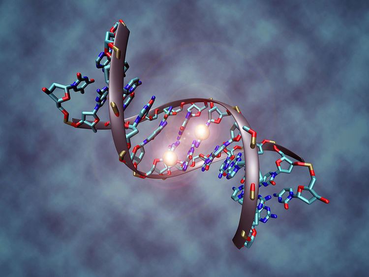
DNA methylation is a process by which methyl groups are added to the DNA molecule. Methylation can change the activity of a DNA segment without changing the sequence. When located in a gene promoter, DNA methylation typically acts to repress gene transcription. DNA methylation is essential for normal development and is associated with a number of key processes including genomic imprinting, X-chromosome inactivation, repression of transposable elements, aging and carcinogenesis.
Contents
- Conserved function of DNA methylation
- CpG islands
- Repression of CpG dense promoters
- Repression of transposable elements
- Methylation of the gene body of highly transcribed genes
- During embryonic development
- In cancer
- In atherosclerosis
- In aging
- In exercise
- In B cell differentiation
- In the brain
- DNA methyltransferases in mammals
- In plants
- In fungi
- In insects
- In bacteria
- Molecular cloning
- Detection
- Differentially methylated regions DMRs
- DNA methylation marks
- Computational prediction
- References
Two of DNA's four bases, cytosine and adenine, can be methylated. Cytosine methylation is widespread in both eukaryotes and prokaryotes, even though the rate of cytosine DNA methylation can differ greatly between species: 14% of cytosines are methylated in Arabidopsis thaliana, 4% in Mus musculus, 2.3% in Escherichia coli, 0.03% in Drosophila, and virtually none (< 0.0002%) in yeast species such as S. cerevisiae and S. pombe (but not N. crassa). Adenine methylation has been observed in bacterial, plant and recently in mammalian DNA, but have received considerably less attention.
Methylation of cytosine to form 5-methylcytosine occurs at the same 5 position on the pyrimidine ring where the DNA base thymine's methyl group is located, distinguishing thymine from the analogous RNA base uracil which has no methyl group. The near-universal replacement of uracil by thymine in DNA, but not RNA, may have evolved as an error-control mechanism, to facilitate removal of uracils generated by the spontaneous deamination of cytosine. DNA methylation as well as many of its contemporary DNA methyltransferases has been thought to evolve from early world primitive RNA methylation activity and is supported by several lines of evidences.
In plants and other organisms, DNA methylation is found in three different sequence contexts: CG (or CpG), CHG or CHH (where H correspond to A,T or C). In mammals however, DNA methylation is almost exclusively found in CpG dinucleotides, with the cytosines on both strand being usually methylated. non-CpG methylation can however be observed in embryonic stem cells, and has also been indicated in neural development. Furthermore, non-CpG methylation has also been observed in hematopoietic progenitor cells, and it occurred mainly in a CpApC sequence context.
Conserved function of DNA methylation
The DNA methylation landscape of vertebrates is very particular compared to other organisms. In vertebrates, around 60-80% of CpG are methylated in somatic cells and DNA methylation appears as a default state that has to be specifically excluded from defined locations. By contrast, the genome of most plants, invertebrates, fungi or protists show “mosaic” methylation patterns, where only specific genomic elements are targeted, and they are characterized by the alternation of methylated and unmethylated domains.
High CpG methylation in mammalian genomes has an evolutionary cost because it increases the frequency of spontaneous mutations. Loss of amino-groups occurs with a high frequency for cytosines, with different consequences depending on their methylation. Methylated C residues spontaneously deaminate to form T residues over time; hence CpG dinucleotides steadily deaminate to TpG dinucleotides, which is evidenced by the under-representation of CpG dinucleotides in the human genome (they occur at only 21% of the expected frequency). (On the other hand, spontaneous deamination of unmethylated C residues gives rise to U residues, a change that is quickly recognized and repaired by the cell.)
CpG islands
In mammals, the only exception for this global CpG depletion resides in a specific category of GC- and CpG-rich sequences termed CpG islands that are generally unmethylated and therefore retained the expected CpG content. CpG islands are usually defined as regions with 1) a length greater than 200bp, 2) a G+C content greater than 50%, 3) a ratio of observed to expected CpG greater than 0.6, although other definitions are sometimes used. Excluding repeated sequences, there are around 25,000 CpG islands in the human genome, 75% of which being less than 850pb long. They are major regulatory units and around 50% of CpG islands are located in gene promoter regions, while another 25% lie in gene bodies, often serving as alternative promoters. Reciprocally, around 60-70% of human genes have a CpG island in their promoter region. The majority of CpG islands are constitutively unmethylated and enriched for permissive chromatin modification such as H3K4 methylation. In somatic tissues, only 10% of CpG islands are methylated, the majority of them being located in intergenic and intragenic regions.
Repression of CpG-dense promoters
DNA methylation was probably present at some extent in very early eukaryote ancestors. In virtually every organism analyzed, methylation in promoter regions correlates negatively with gene expression. CpG-dense promoters of actively transcribed genes are never methylated, but reciprocally transcriptionally silent genes do not necessarily carry a methylated promoter. In mouse and human, around 60-70% of genes have a CpG island in their promoter region and most of these CpG islands remain unmethylated independently of the transcriptional activity of the gene, in both differentiated and undifferentiated cell types. Of note, whereas DNA methylation of CpG islands is unambiguously linked with transcriptional repression, the function of DNA methylation in CG-poor promoters remains unclear; albeit there is little evidence that it could be functionally relevant.
DNA methylation may affect the transcription of genes in two ways. First, the methylation of DNA itself may physically impede the binding of transcriptional proteins to the gene, and second, and likely more important, methylated DNA may be bound by proteins known as methyl-CpG-binding domain proteins (MBDs). MBD proteins then recruit additional proteins to the locus, such as histone deacetylases and other chromatin remodeling proteins that can modify histones, thereby forming compact, inactive chromatin, termed heterochromatin. This link between DNA methylation and chromatin structure is very important. In particular, loss of methyl-CpG-binding protein 2 (MeCP2) has been implicated in Rett syndrome; and methyl-CpG-binding domain protein 2 (MBD2) mediates the transcriptional silencing of hypermethylated genes in cancer.
Repression of transposable elements
DNA methylation is a powerful transcriptional repressor, at least in CpG dense contexts. Transcriptional repression of protein-coding genes appears essentially limited to very specific classes of genes that need to be silent permanently and in almost all tissues. While DNA methylation does not have the flexibility required for the fine-tuning of gene regulation, its stability is perfect to ensure the permanent silencing of transposable elements. Transposon control is one the most ancient function of DNA methylation that is shared by animals, plants and multiple protists. It is even suggested that DNA methylation evolved precisely for this purpose.
Methylation of the gene body of highly transcribed genes
A function that appears even more conserved than transposon silencing is positively correlated with gene expression. In almost all species where DNA methylation is present, DNA methylation is especially enriched in the body of highly transcribed genes. The function of gene body methylation is not well understood. A body of evidence suggests that it could regulate splicing and suppress the activity of intragenic transcriptional units (cryptic promoters or transposable elements). Gene-body methylation appears closely tied to H3K36 methylation. In yeast and mammals, H3K36 methylation is highly enriched in the body of highly transcribed genes. In yeast at least, H3K36me3 recruits enzymes such as histone deacetylases to condense chromatin and prevent the activation of cryptic start sites. In mammals, DNMT3a and DNMT3b PWWP domain binds to H3K36me3 and the two enzymes are recruited to the body of actively transcribed genes.
During embryonic development
DNA methylation patterns are largely erased and then re-established between generations in mammals. Almost all of the methylations from the parents are erased, first during gametogenesis, and again in early embryogenesis, with demethylation and remethylation occurring each time. Demethylation in early embryogenesis occurs in the preimplantation period in two stages – initially in the zygote, then during the first few embryonic replication cycles of morula and blastula. A wave of methylation then takes place during the implantation stage of the embryo, with CpG islands protected from methylation. This results in global repression and allows housekeeping genes to be expressed in all cells. In the post-implantation stage, methylation patterns are stage- and tissue-specific, with changes that would define each individual cell type lasting stably over a long period.
Whereas DNA methylation is not necessary per se for transcriptional silencing, it is thought nonetheless to represent a “locked” state that definitely inactivates transcription. In particular, DNA methylation appears critical for the maintenance of mono-allelic silencing in the context of genomic imprinting and X chromosome inactivation. In these cases, expressed and silent alleles differ by their methylation status, and loss of DNA methylation results in loss of imprinting and re-expression of Xist in somatic cells. During embryonic development, few genes change their methylation status, at the important exception of many genes specifically expressed in the germline. DNA methylation appears absolutely required in differentiated cells, as knockout of any of the three competent DNA methyltransferase results in embryonic or post-partum lethality. By contrast, DNA methylation is dispensable in undifferentiated cell types, such as the inner cell mass of the blastocyst, primordial germ cells or embryonic stem cells. Since DNA methylation appears to directly regulate only a limited number of genes, how precisely DNA methylation absence causes the death of differentiated cells remain an open question.
Due to the phenomenon of genomic imprinting, maternal and paternal genomes are differentially marked and must be properly reprogrammed every time they pass through the germline. Therefore, during gametogenesis, primordial germ cells must have their original biparental DNA methylation patterns erased and re-established based on the sex of the transmitting parent. After fertilization the paternal and maternal genomes are once again demethylated and remethylated (except for differentially methylated regions associated with imprinted genes). This reprogramming is likely required for totipotency of the newly formed embryo and erasure of acquired epigenetic changes.
In cancer
In many disease processes, such as cancer, gene promoter CpG islands acquire abnormal hypermethylation, which results in transcriptional silencing that can be inherited by daughter cells following cell division. Alterations of DNA methylation have been recognized as an important component of cancer development. Hypomethylation, in general, arises earlier and is linked to chromosomal instability and loss of imprinting, whereas hypermethylation is associated with promoters and can arise secondary to gene (oncogene suppressor) silencing, but might be a target for epigenetic therapy.
Global hypomethylation has also been implicated in the development and progression of cancer through different mechanisms. Typically, there is hypermethylation of tumor suppressor genes and hypomethylation of oncogenes.
Generally, in progression to cancer, hundreds of genes are silenced or activated. Although silencing of some genes in cancers occurs by mutation, a large proportion of carcinogenic gene silencing is a result of altered DNA methylation (see DNA methylation in cancer). DNA methylation causing silencing in cancer typically occurs at multiple CpG sites in the CpG islands that are present in the promoters of protein coding genes.
Altered expressions of microRNAs also silence or activate many genes in progression to cancer (see microRNAs in cancer). Altered microRNA expression occurs through hyper/hypo-methylation of CpG sites in CpG islands in promoters controlling transcription of the microRNAs.
Silencing of DNA repair genes through methylation of CpG islands in their promoters appears to be especially important in progression to cancer (see methylation of DNA repair genes in cancer).
In atherosclerosis
Epigenetic modifications such as DNA methylation have been implicated in cardiovascular disease, including atherosclerosis. In animal models of atherosclerosis, vascular tissue as well as blood cells such as mononuclear blood cells exhibit global hypomethylation with gene-specific areas of hypermethylation. DNA methylation polymorphisms may be used as an early biomarker of atherosclerosis since they are present before lesions are observed, which may provide an early tool for detection and risk prevention.
Two of the cell types targeted for DNA methylation polymorphisms are monocytes and lymphocytes, which experience an overall hypomethylation. One proposed mechanism behind this global hypomethylation is elevated homocysteine levels causing hyperhomocysteinemia, a known risk factor for cardiovascular disease. High plasma levels of homocysteine inhibit DNA methyltransferases, which causes hypomethylation. Hypomethylation of DNA affects gene that alter smooth muscle cell proliferation, cause endothelial cell dysfunction, and increase inflammatory mediators, all of which are critical in forming atherosclerotic lesions. High levels of homocysteine also result in hypermethylation of CpG islands in the promoter region of the estrogen receptor alpha (ERα) gene, causing its down regulation. ERα protects against atherosclerosis due to its action as a growth suppressor, causing the smooth muscle cells to remain in a quiescent state. Hypermethylation of the ERα promoter thus allows intimal smooth muscle cells to proliferate excessively and contribute to the development of the atherosclerotic lesion.
Another gene that experiences a change in methylation status in atherosclerosis is the monocarboxylate transporter (MCT3), which produces a protein responsible for the transport of lactate and other ketone bodies out of many cell types, including vascular smooth muscle cells. In atherosclerosis patients, there is an increase in methylation of the CpG islands in exon 2, which decreases MCT3 protein expression. The down regulation of MCT3 impairs lactate transport, and significantly increases smooth muscle cell proliferation, which further contributes to the atherosclerotic lesion. An ex vivo experiment using the demethylating agent Decitabine (5-aza-2 -deoxycytidine) was shown to induce MCT3 expression in a dose dependant manner, as all hypermethylated sites in the exon 2 CpG island became demethylated after treatment. This may serve as a novel therapeutic agent to treat atherosclerosis, although no human studies have been conducted thus far.
In aging
A longitudinal study of twin children showed that, between the ages of 5 and 10, there was divergence of methylation patterns due to environmental rather than genetic influences. There is a global loss of DNA methylation during aging.
In a study that analyzed the complete DNA methylomes of CD4+ T cells in a newborn, a 26 years old individual and a 103 years old individual was observed that the loss of methylation is proportional to age. Hypomethylated CpGs observed in the centenarian DNAs compared with the neonates covered all genomic compartments (promoters, intergenic, intronic and exonic regions).
However, some genes become hypermethylated with age, including genes for the estrogen receptor, p16, and insulin-like growth factor 2. The epigenetic clock is a promising biomarker of aging.
DNA methylation levels can be used to estimate age, forming an accurate biological clock in humans and chimpanzees.
In exercise
High intensity exercise has been shown to result in reduced DNA methylation in skeletal muscle. Promoter methylation of PGC-1α and PDK4 were immediately reduced after high intensity exercise, whereas PPAR-γ methylation was not reduced until three hours after exercise. By contrast, six months of exercise in previously sedentary middle-age men resulted in increased methylation in adipose tissue. One study showed a possible increase in global genomic DNA methylation of white blood cells with more physical activity in non-Hispanics.
In B-cell differentiation
A study that investigated the methylome of B cells along their differentiation cycle, using whole-genome bisulfite sequencing (WGBS), showed that there is a hypomethylation from the earliest stages to the most differentiated stages. The largest methylation difference is between the stages of germinal center B cells and memory B cells. Furthermore, this study showed that there is a similarity between B cell tumors and long-lived B cells in their DNA methylation signatures.
In the brain
Research has suggested that long-term memory storage in humans may be regulated by DNA methylation.
DNA methyltransferases (in mammals)
In mammalian cells, DNA methylation occurs mainly at the C5 position of CpG dinucleotides and is carried out by two general classes of enzymatic activities – maintenance methylation and de novo methylation.
Maintenance methylation activity is necessary to preserve DNA methylation after every cellular DNA replication cycle. Without the DNA methyltransferase (DNMT), the replication machinery itself would produce daughter strands that are unmethylated and, over time, would lead to passive demethylation. DNMT1 is the proposed maintenance methyltransferase that is responsible for copying DNA methylation patterns to the daughter strands during DNA replication. Mouse models with both copies of DNMT1 deleted are embryonic lethal at approximately day 9, due to the requirement of DNMT1 activity for development in mammalian cells.
It is thought that DNMT3a and DNMT3b are the de novo methyltransferases that set up DNA methylation patterns early in development. DNMT3L is a protein that is homologous to the other DNMT3s but has no catalytic activity. Instead, DNMT3L assists the de novo methyltransferases by increasing their ability to bind to DNA and stimulating their activity. Finally, DNMT2 (TRDMT1) has been identified as a DNA methyltransferase homolog, containing all 10 sequence motifs common to all DNA methyltransferases; however, DNMT2 (TRDMT1) does not methylate DNA but instead methylates cytosine-38 in the anticodon loop of aspartic acid transfer RNA.
Since many tumor suppressor genes are silenced by DNA methylation during carcinogenesis, there have been attempts to re-express these genes by inhibiting the DNMTs. 5-Aza-2'-deoxycytidine (decitabine) is a nucleoside analog that inhibits DNMTs by trapping them in a covalent complex on DNA by preventing the β-elimination step of catalysis, thus resulting in the enzymes' degradation. However, for decitabine to be active, it must be incorporated into the genome of the cell, which can cause mutations in the daughter cells if the cell does not die. In addition, decitabine is toxic to the bone marrow, which limits the size of its therapeutic window. These pitfalls have led to the development of antisense RNA therapies that target the DNMTs by degrading their mRNAs and preventing their translation. However, it is currently unclear whether targeting DNMT1 alone is sufficient to reactivate tumor suppressor genes silenced by DNA methylation.
In plants
Significant progress has been made in understanding DNA methylation in the model plant Arabidopsis thaliana. DNA methylation in plants differs from that of mammals: while DNA methylation in mammals mainly occurs on the cytosine nucleotide in a CpG site, in plants the cytosine can be methylated at CpG, CpHpG, and CpHpH sites, where H represents any nucleotide but guanine. Overall, Arabidopsis DNA is highly methylated, mass spectrometry analysis estimated 14% of cytosines to be modified.
The principal Arabidopsis DNA methyltransferase enzymes, which transfer and covalently attach methyl groups onto DNA, are DRM2, MET1, and CMT3. Both the DRM2 and MET1 proteins share significant homology to the mammalian methyltransferases DNMT3 and DNMT1, respectively, whereas the CMT3 protein is unique to the plant kingdom. There are currently two classes of DNA methyltransferases: 1) the de novo class, or enzymes that create new methylation marks on the DNA; and 2) a maintenance class that recognizes the methylation marks on the parental strand of DNA and transfers new methylation to the daughters strands after DNA replication. DRM2 is the only enzyme that has been implicated as a de novo DNA methyltransferase. DRM2 has also been shown, along with MET1 and CMT3 to be involved in maintaining methylation marks through DNA replication. Other DNA methyltransferases are expressed in plants but have no known function (see the Chromatin Database).
It is not clear how the cell determines the locations of de novo DNA methylation, but evidence suggests that, for many (though not all) locations, RNA-directed DNA methylation (RdDM) is involved. In RdDM, specific RNA transcripts are produced from a genomic DNA template, and this RNA forms secondary structures called double-stranded RNA molecules. The double-stranded RNAs, through either the small interfering RNA (siRNA) or microRNA (miRNA) pathways direct de-novo DNA methylation of the original genomic location that produced the RNA. This sort of mechanism is thought to be important in cellular defense against RNA viruses and/or transposons, both of which often form a double-stranded RNA that can be mutagenic to the host genome. By methylating their genomic locations, through an as yet poorly understood mechanism, they are shut off and are no longer active in the cell, protecting the genome from their mutagenic effect. Recently, it was described that methylation of the DNA is the main determinant of embryogenic cultures formation from explants in woody plants and is regarded the main mechanism that explains the poor response of mature explants to somatic embryogenesis in the plants (Isah 2016).
In fungi
Many fungi have low levels (0.1 to 0.5%) of cytosine methylation, whereas other fungi have as much as 5% of the genome methylated. This value seems to vary both among species and among isolates of the same species. There is also evidence that DNA methylation may be involved in state-specific control of gene expression in fungi. However, at a detection limit of 250 attomoles by using ultra-high sensitive mass spectrometry DNA methylation was not confirmed in single cellular yeast species such as Saccharomyces cerevisiae or Schizosaccharomyces pombe, indicating that yeasts do not possess this DNA modification.
Although brewers' yeast (Saccharomyces), fission yeast (Schizosaccharomyces), and Aspergillus flavus have no detectable DNA methylation, the model filamentous fungus Neurospora crassa has a well-characterized methylation system. Several genes control methylation in Neurospora and mutation of the DNA methyl transferase, dim-2, eliminates all DNA methylation but does not affect growth or sexual reproduction. While the Neurospora genome has very little repeated DNA, half of the methylation occurs in repeated DNA including transposon relics and centromeric DNA. The ability to evaluate other important phenomena in a DNA methylase-deficient genetic background makes Neurospora an important system in which to study DNA methylation.
In insects
Functional DNA methylation has been discovered in Honey Bees. DNA methylation marks are mainly on the gene body, and current opinions on the function of DNA methylation is gene regulation via alternative splicing
Drosophila melanogaster possess a very low level of DNA methylation only that is however too low to be studied by methods such as bisulphite sequencing. Takayama et al. developed a sensitive method that allowed to find that the fly genome DNA sequence patterns that associate with methylation are very different from the patterns seen in humans, or in other animal or plant species to date. Genome methylation in D. melanogaster was found at specific short motifs (concentrated in specific 5-base sequence motifs that are CA- and CT-rich but depleted of guanine) and is independent of DNMT2 activity. By using highly sensitive mass spectrometry approaches, Zhang et al. have now demonstrated the presence of low (0.07%) but significant levels of adenine methylation during the earliest stages of Drosophila embryogenesis.
In bacteria
Adenine or cytosine methylation is part of the restriction modification system of many bacteria, in which specific DNA sequences are methylated periodically throughout the genome. A methylase is the enzyme that recognizes a specific sequence and methylates one of the bases in or near that sequence. Foreign DNAs (which are not methylated in this manner) that are introduced into the cell are degraded by sequence-specific restriction enzymes and cleaved. Bacterial genomic DNA is not recognized by these restriction enzymes. The methylation of native DNA acts as a sort of primitive immune system, allowing the bacteria to protect themselves from infection by bacteriophage.
E. coli DNA adenine methyltransferase (Dam) is an enzyme of ~32 kDa that does not belong to a restriction/modification system. The target recognition sequence for E. coli Dam is GATC, as the methylation occurs at the N6 position of the adenine in this sequence (G meATC). The three base pairs flanking each side of this site also influence DNA–Dam binding. Dam plays several key roles in bacterial processes, including mismatch repair, the timing of DNA replication, and gene expression. As a result of DNA replication, the status of GATC sites in the E. coli genome changes from fully methylated to hemimethylated. This is because adenine introduced into the new DNA strand is unmethylated. Re-methylation occurs within two to four seconds, during which time replication errors in the new strand are repaired. Methylation, or its absence, is the marker that allows the repair apparatus of the cell to differentiate between the template and nascent strands. It has been shown that altering Dam activity in bacteria results in increased spontaneous mutation rate. Bacterial viability is compromised in dam mutants that also lack certain other DNA repair enzymes, providing further evidence for the role of Dam in DNA repair.
One region of the DNA that keeps its hemimethylated status for longer is the origin of replication, which has an abundance of GATC sites. This is central to the bacterial mechanism for timing DNA replication. SeqA binds to the origin of replication, sequestering it and thus preventing methylation. Because hemimethylated origins of replication are inactive, this mechanism limits DNA replication to once per cell cycle.
Expression of certain genes, for example those coding for pilus expression in E. coli, is regulated by the methylation of GATC sites in the promoter region of the gene operon. The cells' environmental conditions just after DNA replication determine whether Dam is blocked from methylating a region proximal to or distal from the promoter region. Once the pattern of methylation has been created, the pilus gene transcription is locked in the on or off position until the DNA is again replicated. In E. coli, these pilus operons have important roles in virulence in urinary tract infections. It has been proposed that inhibitors of Dam may function as antibiotics.
On the other hand, DNA cytosine methylase targets CCAGG and CCTGG sites to methylate cytosine at the C5 position (C meC(A/T) GG). The other methylase enzyme, EcoKI, causes methylation of adenines in the sequences AAC(N6)GTGC and GCAC(N6)GTT.
Molecular cloning
Most strains used by molecular biologists are derivatives of E. coli K-12, and possess both Dam and Dcm, but there are commercially available strains that are dam-/dcm- (lack of activity of either methylase). In fact, it is possible to unmethylate the DNA extracted from dam+/dcm+ strains by transforming it into dam-/dcm- strains. This would help digest sequences that are not being recognized by methylation-sensitive restriction enzymes.
The restriction enzyme DpnI can recognize 5'-GmeATC-3' sites and digest the methylated DNA. Being such a short motif, it occurs frequently in sequences by chance, and as such its primary use for researchers is to degrade template DNA following PCRs (PCR products lack methylation, as no methylases are present in the reaction). Similarly, some commercially available restriction enzymes are sensitive to methylation at their cognate restriction sites, and must as mentioned previously be used on DNA passed through a dam-/dcm- strain to allow cutting.
Detection
DNA methylation can be detected by the following assays currently used in scientific research:
Differentially methylated regions (DMRs)
Differentially methylated regions, are genomic regions with different methylation statuses among multiple samples (tissues, cells, individuals or others), are regarded as possible functional regions involved in gene transcriptional regulation. The identification of DMRs among multiple tissues (T-DMRs) provides a comprehensive survey of epigenetic differences among human tissues. For example, these methylated regions that are unique to a particular tissue allow individuals to differentiate between tissue type, such as semen and vaginal fluid. Current research conducted by Lee et al., showed DACT1 and USP49 positively identified semen by examining T-DMRs. DMRs between cancer and normal samples (C-DMRs) demonstrate the aberrant methylation in cancers. It is well known that DNA methylation is associated with cell differentiation and proliferation. Many DMRs have been found in the development stages (D-DMRs) and in the reprogrammed progress (R-DMRs). In addition, there are intra-individual DMRs (Intra-DMRs) with longitudinal changes in global DNA methylation along with the increase of age in a given individual. There are also inter-individual DMRs (Inter-DMRs) with different methylation patterns among multiple individuals.
QDMR (Quantitative Differentially Methylated Regions) is a quantitative approach to quantify methylation difference and identify DMRs from genome-wide methylation profiles by adapting Shannon entropy <http://bioinfo.hrbmu.edu.cn/qdmr>. The platform-free and species-free nature of QDMR makes it potentially applicable to various methylation data. This approach provides an effective tool for the high-throughput identification of the functional regions involved in epigenetic regulation. QDMR can be used as an effective tool for the quantification of methylation difference and identification of DMRs across multiple samples.
Gene-set analysis (a.k.a. pathway analysis; usually performed tools such as DAVID, GoSeq or GSEA) has been shown to be severely biased when applied to high-throughput methylation data (e.g. MeDIP-seq, MeDIP-ChIP, HELP-seq etc.), and a wide range of studies have thus mistakenly reported hyper-methylation of genes related to development and differentiation; it has been suggested that this can be corrected using sample label permutations or using a statistical model to control for differences in the numberes of CpG probes / CpG sites that target each gene.
DNA methylation marks
DNA methylation marks, are genomic regions with specific methylation pattern in specific biological state such as tissue, cell type, individual), are regarded as possible functional regions involved in gene transcriptional regulation. Although various human cell types may have the same genome, these cells have different methylomes. The systematic identification and characterization of methylation marks across cell types are crucial to understand the complex regulatory network for cell fate determination. Hongbo Liu et al. proposed an entropy-based framework termed SMART to integrate the whole genome bisulfite sequencing methylomes across 42 human tissues/cells and identified 757,887 genome segments. Nearly 75% of the segments showed uniform methylation across all cell types. From the remaining 25% of the segments, they identified cell type-specific hypo/hypermethylation marks that were specifically hypo/hypermethylated in a minority of cell types using a statistical approach and presented an atlas of the human methylation marks. Further analysis revealed that the cell type-specific hypomethylation marks were enriched through H3K27ac and transcription factor binding sites in cell type-specific manner. In particular, they observed that the cell type-specific hypomethylation marks are associated with the cell type-specific super-enhancers that drive the expression of cell identity genes. This framework provides a complementary, functional annotation of the human genome and helps to elucidate the critical features and functions of cell type-specific hypomethylation.
The entropy-based Specific Methylation Analysis and Report Tool, termed "SMART", which focus on integrating a large number of DNA methylomes for the de novo identification of cell type-specific methylation marks.
Computational prediction
DNA methylation can also be detected by computational models through sophisticated algorithms and methods. Computational models can facilitate the global profiling of DNA methylation across chromosomes, and often such models are faster and cheaper to perform than biological assays. Such up-to-date computational models include Bhasin, et al., Bock, et al., and Zheng, et al. Together with biological assay, these methods greatly facilitate the DNA methylation analysis.