
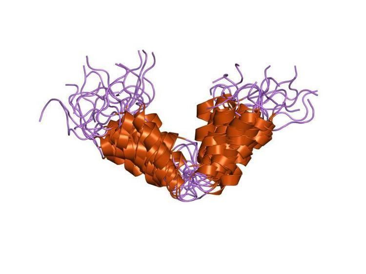
Symbol P19Arf_N InterPro IPR010868 SUPERFAMILY 1hn3 | Pfam PF07392 SCOP 1hn3 Pfam structures | |
 | ||
p16 (also known as cyclin-dependent kinase inhibitor 2A, multiple tumor suppressor 1 and as several other synonyms), is a tumor suppressor protein, that in humans is encoded by the CDKN2A gene. p16 plays an important role in cell cycle regulation by decelerating cells progression from G1 phase to S phase, and therefore acts as a tumor suppressor that is implicated in the prevention of cancers, notably melanoma, oropharyngeal squamous cell carcinoma, cervical cancer, and esophageal cancer. p16 can be used to improve the histological diagnostic accuracy of CIN3. The CDKN2A gene is frequently mutated or deleted in a wide variety of tumors.
Contents
- Nomenclature
- Gene
- Function
- Regulation
- Role in cancer
- Use as a biomarker
- p16 FISH
- gynecologic cancers
- Urinary bladder SCCs
- Role in senescence
- Experimental analysis of p16 mutation
- Discovery
- Interactions
- References
p16 is an inhibitor of cyclin dependent kinases such as CDK4 and CDK6. These latter kinases phosphorylate retinoblastoma protein (pRB) which eventually results in progression from G1 phase to S phase.
p16 was originally found in an “open reading frame of 148 amino acids encoding a protein of molecular weight 15,845 comprising four ankyrin repeats.” p16Ink4A is named after its molecular weight and its role in inhibiting CDK4.
Nomenclature
p16 is also known as:
Gene
In humans, p16 is encoded by the CDKN2A gene, located on chromosome 9 (9p21.3). This gene generates several transcript variants that differ in their first exons. At least three alternatively spliced variants encoding distinct proteins have been reported, two of which encode structurally related isoforms known to function as inhibitors of CDK4. The remaining transcript includes an alternate exon 1 located 20 kb upstream of the remainder of the gene; this transcript contains an alternate open reading frame (ARF) that specifies a protein that is structurally unrelated to the products of the other variants. The ARF product functions as a stabilizer of the tumor suppressor protein p53, as it can interact with and sequester MDM2, a protein responsible for the degradation of p53. In spite of their structural and functional differences, the CDK inhibitor isoforms and the ARF product encoded by this gene, through the regulatory roles of CDK4 and p53 in cell cycle G1 progression, share a common functionality in control of the G1 phase of the cell cycle. This gene is frequently mutated or deleted in a wide variety of tumors and is known to be an important tumor suppressor gene.
Increased expression of the p16 gene as organisms age reduces the proliferation of stem cells. This reduction in the division and production of stem cells protects against cancer while increasing the risks associated with cellular senescence.
Function
p16 is a cyclin-dependent kinase (CDK) inhibitor that slows down the cell cycle by prohibiting progression from G1 phase to S phase. Normally, CDK4/6 binds cyclin D and forms an active protein complex that phosphorylates retinoblastoma protein (pRB). Once phosphorylated, pRB disassociates from the transcription factor E2F1, liberating E2F1 from its cytoplasm bound state allowing it to enter the nucleus. Once in the nucleus, E2F1 promotes the transcription of target genes that are essential for transition from G1 to S phase.
p16 acts as a tumor suppressor by binding to CDK4/6 and preventing its interaction with cyclin D. This interaction ultimately inhibits the downstream activities of transcription factors, such as E2F1, and arrests cell proliferation. This pathway connects the processes of tumor oncogenesis and senescence, fixing them on opposite ends of a spectrum. On one end, the hypermethylation, mutation, or deletion of p16 leads to downregulation of the gene and can lead to cancer through the dysregulation of cell cycle progression. Conversely, activation of p16 through the ROS pathway, DNA damage, or senescence leads to the buildup of p16 in tissues and is implicated in aging of cells.
Regulation
Regulation of p16 is complex and involves the interaction of several transcription factors, as well as several proteins involved in epigenetic modification through methylation and repression of the promoter region.
PRC1 and PRC2 are two protein complexes that modify the expression of p16 through the interaction of various transcription factors that execute methylation patterns that can repress transcription of p16. These pathways are activated in cellular response to reduce senescence.
Role in cancer
Mutations resulting in deletion or reduction of function of the CDKN2A gene are associated with increased risk of a wide range of cancers and alterations of the gene are frequently seen in cancer cell lines. Examples include:
Pancreatic adenocarcinoma is often associated with mutations in the CDKN2A gene.
Carriers of germline mutations in CDKN2A have besides their high risks of melanoma also increased risks of pancreatic, lung, laryngeal and oropharyngeal cancers and tobacco smoking exacerbates carriers’ susceptibility for such non-melanoma cancers.
Homozygous deletion of p16 are frequently found in esophageal cancer and gastric cancer cell lines.
Germline mutations in CDKN2A are associated with an increased susceptibility to develop skin cancer.
Hypermethylation of tumor suppressor genes has been implicated in various cancers. In 2013, a meta-analysis of 39 articles using analysis cancer tissues and 7 articles using blood samples, revealed an increased frequency of DNA methylation of p16 gene in esophageal cancer. As the degree of tumor differentiation increased, so did the frequency of DNA methylation.
Tissue samples of primary oral squamous cell carcinoma (OSCC) display hypermethylation in the promoter regions of p16. Cancer cells show a significant increase in the accumulation of methylation in CpG islands in the promoter region of p16. This epigenetic change leads to the loss of tumor suppressor gene function through two possible mechanisms. Methylation can physically inhibit the transcription of the gene or methylation can lead to the recruitment of transcription factors that repress transcription. Both mechanisms lead to the same end result—downregulation of gene expression that leads to decreased levels of the p16 protein. It has been suggested that this process is responsible for the development of various forms of cancer serving as an alternative process to gene deletion or mutation.
Use as a biomarker
Furthermore, p16 is now being explored as a prognostic biomarker for a number of cancers. For patients with oropharyngeal squamous cell carcinoma, using immunohistochemistry to detect the presence of the p16 biomarker has been shown to be the strongest indicator of disease course. Presence of the biomarker is associated with a more favorable prognosis as measured by cancer-specific survival (CSS), recurrence-free survival (RFS), locoregional control (LRC), as well as other measurements. The appearance of hyper methylation of p16 is also being evaluated as a potential prognostic biomarker for prostate cancer.
p16 FISH
p16 deletion detected by FISH in surface epithelial mesothelial proliferations is predictive of underlying invasive mesothelioma.
gynecologic cancers
p16 is a widely used immunohistochemical marker in gynecologic pathology. Strong and diffuse cytoplasmic and nuclear expression of p16 in squamous cell carcinomas (SCC) of the female genital tract is strongly associated with high-risk human papilloma virus (HPV) infection and neoplasms of cervical origin. The majority of SCCs of uterine cervix express p16. However, p16 can be expressed in other neoplasms and in several normal human tissues.
Urinary bladder SCCs
More than a third of urinary bladder SCCs express p16. SCCs of urinary bladder express p16 independent of gender. p16 immunohistochemical expression alone cannot be used to discriminate between SCCs arising from uterine cervix versus urinary bladder.
Role in senescence
Concentrations of p16INK4a increase dramatically as tissue ages. p16INK4a, along with senescence-associated beta-galactosidase, is regarded to be a biomarker of cellular senescence. Therefore, p16INK4a could potentially be used as a blood test that measures how fast the body's tissues are aging at a molecular level.
It has been used as a target to delay some aging changes in mice.
Experimental analysis of p16 mutation
As consensus grows regarding the strength of p16 as a biomarker for detecting and determining prognoses of cancer, p16 immunohistochemistry is growing in importance.
Discovery
Researchers Manuel Serrano, Gregory J. Hannon and David Beach discovered p16 in 1993 and correctly characterized the protein as a cyclin-dependent kinase inhibitor. Since its discovery, p16 has become significant in the field of cancer research. The protein was suspected to be involved in carcinogenesis due to the observation that mutation or deletion in the gene was implicated in human cancer cell lines. The detection of p16 inactivation in familial melanoma supplied further evidence. p16 deletion, mutation, or hypermethylation is now associated with various cancers. Whether p16 can be considered to be a driver mutation requires further investigation.
Interactions
P16 (gene) has been shown to interact with: