
Specialty gastroenterology | ||
 | ||
ICD-10 K04.8, K09.1, K09.2, K09.8, K09.9 and K10.0 | ||
A cyst is a pathological epithelial lined cavity that fills with fluid or soft material and usually grows from internal pressure generated by fluid being drawn into the cavity from osmosis (hydrostatic pressure). The bones of the jaws, the mandible and maxilla, are the bones with the highest prevalence of cysts in the human body. This is due to the abundant amount of epithelial remnants that can be left in the bones of the jaws. The enamel of teeth is formed from ectoderm (the precursor germ layer to skin and mucosa), and so remnants of epithelium can be left in the bone during odontogenesis (tooth development). The bones of the jaws develop from embryologic processes which fuse together, and ectodermal tissue may be trapped along the lines of this fusion. This "resting" epithelium (also termed cell rests) is usually dormant or undergoes atrophy, but, when stimulated, may form a cyst. The reasons why resting epithelium may proliferate and undergo cystic transformation are generally unknown, but inflammation is thought to be a major factor. The high prevalence of tooth impactions and dental infections that occur in the bones of the jaws is also significant to explain why cysts are more common at these sites.
Contents
- Odontogenic cysts
- Developmental Non odontogenic cysts
- Developmental cysts of the jaws
- Developmental cysts of the soft tissues around the jaws
- Developmental cysts of questionable etiology
- Signs and symptoms
- Diagnosis
- Treatment
- Prognosis
- Epidemiology
- References
Cysts that arise from tissue(s) that would normally develop into teeth are referred to as odontogenic cysts. Other cysts of the jaws are termed non-odontogenic cysts. Non-odontogenic cysts form from tissues other than those involved in tooth development, and consequently may contain structures such as epithelium from the nose. As the cyst grows from hydraulic pressure it causes the bone around it to resorb, and may cause movement of teeth or other vital structures such as nerves and blood vessels, or resorb the roots of teeth. Most cysts do not cause any symptoms, and are discovered on routine dental radiographs. Some cysts may not require any treatment, but if treatment is required, it usually involves some minor surgery to partially or completely remove the cyst in a one or two-stage procedure.
Odontogenic cysts
Odontogenic cysts have histologic origins in the cells of the dental structures. Some are inflammatory while others are developmental.
Developmental/ Non-odontogenic cysts
There are several development cysts of the head and neck most of which form in the soft tissues rather than the bone. There are also several cysts, previously thought to arise from epithelial remanents trapped in embryonic lines of fusion, most of which are now believed to be odontogenic in origin or have an unknown etiology. Their names are included for the sake of completeness.
Developmental cysts of the jaws
Developmental cysts of the soft tissues around the jaws
Developmental cysts of questionable etiology
Signs and symptoms
Cysts rarely cause any symptoms, unless they become secondarily infected. The signs depend mostly upon the size and location of the cyst. If the cyst has not expanded beyond the normal anatomical boundaries of the bone, then there will be no palpable lump outside or inside the mouth. The vast majority of cysts expand slowly, and the surrounding bone has time to increase its density around the lesion, which is the body's attempt to isolate the lesion. Cysts that have expanded beyond the normal anatomic boundaries of a bone are still often covered with a thin layer of new bone. At this stage, there may be a sign termed "eggshell cracking", where the thinned cortical plate cracks when pressure is applied. A lump may be felt, which may feel hard if there is still bone covering the cyst, or fluctuant if the cyst has eroded through the bone surrounding it. A cyst may become acutely infected, and discharge into the oral cavity via a sinus. Adjacent teeth may be loosened, tilted or even moved bodily. Rarely, roots of teeth are resorbed, depending upon the type of cyst. The inferior alveolar nerve runs through the mandible and supplies sensation to the lower lip and chin. As most cysts expand slowly, there will be no altered sensation (anesthesia or paraesthesia), since the inferior alveolar canal is harmlessly enveloped or displaced over time. More aggressive cysts, or acute infection of any cyst may cause altered sensation.
Diagnosis
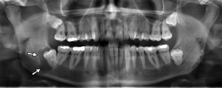
Most cysts are discovered as a chance finding on routine dental radiography. On an x-ray, cysts appear as radiolucent (dark) areas with radiopaque (white) borders. Cysts are usually unilocular, but may also be multilocular. Sometimes aspiration is used to aid diagnosis of a cystic lesion, e.g. fluid aspirate from a radicular cyst may appear straw colored and display shimmering due to cholesterol content. Almost always, the cyst lining is sent to a pathologist for histopathologic examination after it has been surgically removed. This means that the exact diagnosis of the type of cyst is often made in retrospect.
Treatment
When treatment is required, this is usually by surgical removal of the cyst. There are four ways in which cysts are managed:
Prognosis
The prognosis depends upon the type, size and location of a cyst. Most cysts are entirely benign, and some may require no treatment. Rarely, some cystic lesions represent locally aggressive tumors that may cause destruction of surrounding bone if left untreated. This type of cyst are usually removed with a margin of healthy bone to prevent recurrence of new cysts. If a cyst expands to a very large size, the mandible may be weakened such that a pathologic fracture occurs.
Epidemiology
Radicular cysts are by far the most common cyst occurring in the jaws.