
 | ||
Molecular binding is an interaction between molecules that results in a stable physical association between those molecules. Cooperative binding occurs in binding systems containing more than one type, or species, of molecule and in which one of the partners is not mono-valent and can bind more than one molecule of the other species.
Contents
- Christian Bohr and the concept of cooperative binding
- The Hill equation
- The Adair equation
- The Klotz equation
- Pauling equation
- The KNF model
- The MWC model
- Examples
- Multimeric enzymes
- Ion channels
- Multi site molecules
- Transcription factors
- Conformational spread and binding cooperativity
- References
For example, consider a system where one molecule of species A can bind two molecules of species B. Species A is called the receptor and species B is called the ligand. Binding can be considered "cooperative" if the binding of the first molecule of B to A changes the binding affinity of the second B molecule, making it more or less likely to bind. In other words, the binding of B molecules to the different sites on A do not constitute mutually independent events.
Cooperativity can be positive or negative. Cooperative binding is observed in many biopolymers, including proteins and nucleic acids. Cooperative binding has been shown to be the mechanism underlying a large range of biochemical and physiological processes.
Christian Bohr and the concept of cooperative binding
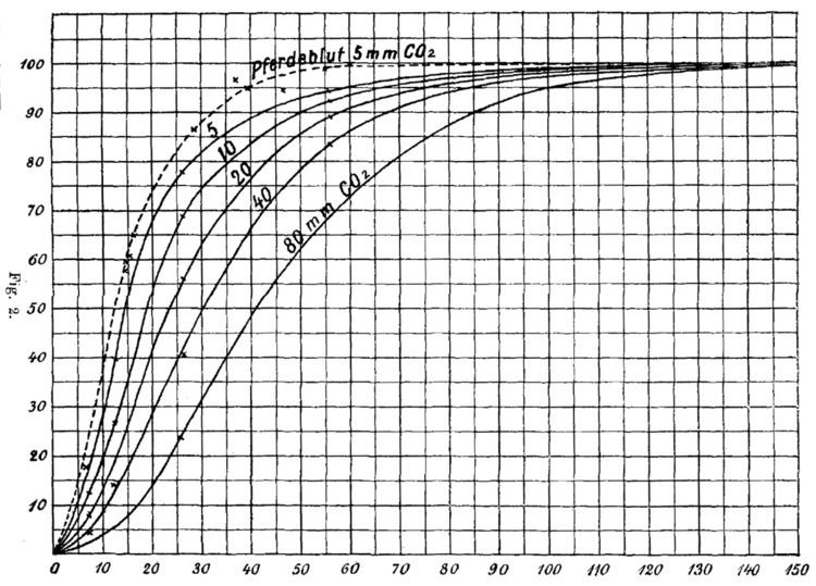
In 1904, Christian Bohr studied hemoglobin binding to oxygen under different conditions. When plotting hemoglobin saturation with oxygen as a function of the partial pressure of oxygen, he obtained a sigmoidal (or "S-shaped") curve, see Figure 1. This indicates that the more oxygen is bound to hemoglobin, the easier it is for more oxygen to bind - until all binding sites are saturated. In addition, Bohr noticed that increasing CO2 pressure shifted this curve to the right - i.e. higher concentrations of CO2 make it more difficult for hemoglobin to bind oxygen. This latter phenomenon, together with the observation that hemoglobin's affinity for oxygen increases with increasing pH, is known as the Bohr effect.
A receptor molecule is said to exhibit cooperative binding if its binding to ligand scales non-linearly with ligand concentration. Cooperativity can be positive (if binding of a ligand molecule increases the receptor's apparent affinity, and hence increases the chance of another ligand molecule binding) or negative (if binding of a ligand molecule decreases affinity and hence makes binding of other ligand molecules less likely). Figure 1 is a chart of the "fractional occupancy"
If
The concept of cooperative binding only applies to molecules or complexes with more than one ligand binding sites. If several ligand binding sites exist, but ligand binding to any one site does not affect the others, the receptor is said to be non-cooperative. Cooperativity can be homotropic, if a ligand influences the binding of ligands of the same kind, or heterotropic, if it influences binding of other kinds of ligands. In the case of hemoglobin, Bohr observed homotropic positive cooperativity (binding of oxygen facilitates binding of more oxygen) and heterotropic negative cooperativity (binding of CO2 reduces hemoglobin's facility to bind oxygen.)
Throughout the 20th century, various frameworks have been developed to describe the binding of a ligand to a protein with more than one binding site and the cooperative effects observed in this context (reviewed by Wyman, J. and Gill, 1990).
The Hill equation
The first description of cooperative binding to a multi-site protein was developed by A.V. Hill. Drawing on observations of oxygen binding to hemoglobin and the idea that cooperativity arose from the aggregation of hemoglobin molecules, each one binding one oxygen molecule, Hill suggested a phenomenological equation that has since been named after him:
where
The "Hill plot" is obtained by plotting
The Adair equation
G.S. Adair found that the Hill plot for hemoglobin was not a straight line, and hypothesized that binding affinity was not a fixed term, but dependent on ligand saturation. Having demonstrated that hemoglobin contained four hemes (and therefore binding sites for oxygen), he worked from the assumption that fully saturated hemoglobin is formed in stages, with intermediate forms with one, two, or three bound oxygen molecules. The formation of each intermediate stage from unbound hemoglobin can be described using an apparent macroscopic association constant
Or, for any protein with n ligand binding sites:
where n denotes the number of binding sites and each
The Klotz equation
Working on calcium binding proteins, Irving Klotz deconvoluted Adair's association constants by considering stepwise formation of the intermediate stages, and tried to express the cooperative binding in terms of elementary processes governed by mass action law. In his framework,
It is worth noting that the constants
The Klotz equation (which is sometimes also called the Adair-Klotz equation) is still often used in the experimental literature to describe measurements of ligand binding in terms of sequential apparent binding constants.
Pauling equation
By the middle of the 20th century, there was an increased interest in models that would not only describe binding curves phenomenologically, but offer an underlying biochemical mechanism. Linus Pauling reinterpreted the equation provided by Adair, assuming that his constants were the combination of the binding constant for the ligand (
The KNF model
Based on results showing that the structure of cooperative proteins changed upon binding to their ligand, Daniel Koshland and colleagues refined the biochemical explanation of the mechanism described by Pauling. The Koshland-Némethy-Filmer (KNF) model assumes that each subunit can exist in one of two conformations: active or inactive. Ligand binding to one subunit would induce an immediate conformational change of that subunit from the inactive to the active conformation, a mechanism described as "induced fit". Cooperativity, according to the KNF model, would arise from interactions between the subunits, the strength of which varies depending on the relative conformations of the subunits involved. For a tetrahedric structure (they also considered linear and square structures), they proposed the following formula:
Where
The MWC model
The Monod-Wyman-Changeux (MWC) model for concerted allosteric transitions went a step further by exploring cooperativity based on thermodynamics and three-dimensional conformations. It was originally formulated for oligomeric proteins with symmetrically arranged, identical subunits, each of which has one ligand binding site. According to this framework, two (or more) interconvertible conformational states of an allosteric protein coexist in a thermal equilibrium. The states - often termed tense (T) and relaxed (R) - differ in affinity for the ligand molecule. The ratio between the two states is regulated by the binding of ligand molecules that stabilizes the higher-affinity state. Importantly, all subunits of a molecule change states at the same time, a phenomenon known as "concerted transition". The MWC model is illustrated in Figure 3.
The allosteric isomerisation constant L describes the equilibrium between both states when no ligand molecule is bound:
The sigmoid Hill plot of allosteric proteins (shown in Figure 5) can then be analysed as a progressive transition from the T state (low affinity) to the R state (high affinity) as the saturation increases (see Figure 4). The slope of the Hill plot also depends on saturation, with a maximum value at the inflexion point. The intercepts between the two asymptotes and the y-axis allow to determine the affinities of both states for the ligand.
In proteins, conformational change is often associated with activity, or activity towards specific targets. Such activity is often what is physiologically relevant or what is experimentally measured. The degree of conformational change is described by the state function
A crucial aspect of the MWC model is that the curves for
If an allosteric protein binds to a target that also has a higher affinity for the R state, then target binding further stabilizes the R state, hence increasing ligand affinity. If, on the other hand, a target preferentially binds to the T state, then target binding will have a negative effect on ligand affinity. Such targets are called allosteric modulators.
Since its inception, the MWC framework has been extended and generalized. Variations have been proposed, for example to cater for proteins with more than two states, proteins that bind to several types of ligands or several types of allosteric modulators and proteins with non-identical subunits or ligand-binding sites.
Examples
The list of molecular assemblies that exhibit cooperative binding of ligands is very large, but some examples are particularly notable for their historical interest, their unusual properties, or their physiological importance.
As described in the historical section, the most famous example of cooperative binding is hemoglobin. Its quaternary structure, solved by Max Perutz using X-ray diffraction, exhibits a pseudo-symmetrical tetrahedron carrying four binding sites (hemes) for oxygen (see Figure 6). Many other molecular assemblies exhibiting cooperative binding have been studied in great detail.
Multimeric enzymes
The activity of many enzymes is regulated by allosteric effectors. Some of these enzymes are multimeric and carry several binding sites for the regulators.
Threonine deaminase was one of the first enzymes suggested to behave like hemoglobin and shown to bind ligands cooperatively. It was later shown to be a tetrameric protein.
Another enzyme that has been suggested early to bind ligands cooperatively is aspartate trans-carbamylase. Although initial models were consistent with four binding sites, its structure was later shown to be hexameric by William Lipscomb and colleagues.
Ion channels
Most ion channels are formed of several identical or pseudo-identical monomers or domains, arranged symmetrically in biological membranes. Several classes of such channels whose opening is regulated by ligands exhibit cooperative binding of these ligands.
It was suggested as early as 1967 (when the exact nature of those channels was still unknown) that the nicotinic acetylcholine receptors bound acetylcholine in a cooperative manner due to the existence of several binding sites. The purification of the receptor and its characterization demonstrated a pentameric structure with binding sites located at the interfaces between subunits, confirmed by the structure of the receptor binding domain.
Inositol triphosphate (IP3) receptors form another class of ligand-gated ion channels exhibiting cooperative binding. The structure of those receptors shows four IP3 binding sites symmetrically arranged.
Multi-site molecules
Although most proteins showing cooperative binding are multimeric complexes of homologous subunits, some proteins carry several binding sites for the same ligand on the same polypeptide. One such example is calmodulin. One molecule of calmodulin binds four calcium ions cooperatively. Its structure presents four EF-hand domains, each one binding one calcium ion. Interestingly, the molecule does not display a square or tetrahedron structure, but is formed of two lobes, each carrying two EF-hand domains.
Transcription factors
Cooperative binding of proteins onto nucleic acids has also been shown. A classical example is the binding of the lambda phage repressor to its operators, which occurs cooperatively. Other examples of transcription factors exhibit positive cooperativity when binding their target, such as the repressor of the TtgABC pumps (n=1.6).
Conversely, examples of negative cooperativity for the binding of transcription factors were also documented, as for the homodimeric repressor of the Pseudomonas putida cytochrome P450cam hydroxylase operon (n=0.56).
Conformational spread and binding cooperativity
Early on, it has been argued that some proteins, especially those consisting of many subunits, could be regulated by a generalized MWC mechanism, in which the transition between R and T state is not necessarily synchronized across the entire protein. In 1969, Wyman proposed such a model with "mixed conformations" (i.e. some protomers in the R state, some in the T state) for respiratory proteins in invertebrates.
Following a similar idea, the conformational spread model by Duke and colleagues subsumes both the KNF and the MWC model as special cases. In this model, a subunit does not automatically change conformation upon ligand binding (as in the KNF model), nor do all subunits in a complex change conformations together (as in the MWC model). Conformational changes are stochastic with the likelihood of a subunit switching states depending on whether or not it is ligand bound and on the conformational state of neighbouring subunits. Thus, conformational states can "spread" around the entire complex.