
Specialty medical genetics ICD-9-CM 728.3, 754.89 eMedicine ped/142 | ICD-10 Q74.3 DiseasesDB 31688 | |
 | ||
OMIM 108110 108120 208100 301830 601701 208200 108200 301830 208155 601680 108145 208085 | ||
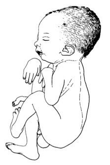
Arthrogryposis multiplex congenita (AMC), or simply arthrogryposis, describes congenital joint contractures in two or more areas of the body. It derives its name from Greek, literally meaning "curving of joints" (arthron, "joint"; grȳpōsis, late Latin form of late Greek grūpōsis, "hooking"). Children born with one or more joint contractures have abnormal fibrosis of the muscle tissue causing muscle shortening, and therefore are unable to perform active extension and flexion in the affected joint or joints. AMC has been divided into three groups: amyoplasia, distal arthrogryposis, and syndromic. Amyoplasia is characterized by severe joint contractures and muscle weakness. Distal arthrogryposis mainly involves the hands and feet. Types of arthrogryposis with a primary neurological or muscle disease belong to the syndromic group.
Contents
Signs and symptoms
Almost every joint in a patient with arthrogryposis is often affected; in 84% all limbs are involved, in 11% only the legs, and in 4% only the arms are involved. Every joint in the body has typical signs and symptoms like the shoulder (internal rotation), wrist (volar and ulnar), hand (fingers in fixed flexion and thumb in palm), hip (flexed, abducted and externally rotated, frequently dislocated), elbow (extension and pronation) and foot (clubfoot). The range of motion capability can be different between joints because of the different deviations. Some types of arthrogryposis like amyoplasia have a symmetrical joint/limb involvement, with normal sensations. The contractures in the joints are sometimes resulting in a reduced walking development in the first 5 years. The intelligence is normal to above normal in children with amyoplasia. But it is unknown how many of these children have an above normal intelligence and there is no literature available about the cause of this syndrome. There are a few syndromes like the Freeman-Sheldon and Gordon syndrome, which have craniofacial involvement. The amyoplasia form of arthrogryposis is sometimes accompanied with a midline facial hemangioma. Arthrogryposis is not a diagnosis but a clinical finding. So this disease is often accompanied with other syndromes or diseases. These other diagnoses can be found in every single organ in a patient. There are a few slightly more common diagnoses such as pulmonary hypoplasia, cryptorchidism, congenital heart defects, tracheoesophageal fistulas, inguinal hernias, cleft palate, and eye abnormalities.
Causes
Research of arthrogryposis has shown that anything that inhibits normal joint movement before birth can result in joint contractures. Arthrogryposis could be caused by genetic and environmental factors. In principle: any factor that curtails fetal movement can result to congenital contractures. The exact causes of arthrogryposis are unknown yet.
Extrinsic factors
The malformations of arthrogryposis can be secondary to environmental factors such as: decreased intrauterine movement, oligohydramnios (low volume or abnormal distribution of intrauterine fluid), and defects in the fetal blood supply. Other causes could be: hyperthermia, limb immobilization and viral infections. Myasthenia gravis of the mother leads also in rare cases to arthrogryposis. The major cause in humans is fetal akinesia. However, this is disputed lately.
Intrinsic factors
Arthrogryposis could also be caused by intrinsic factors. This includes molecular, muscle- and connective tissue development disorders or neurological abnormalities.
Molecular basis
Research has shown that there are more than 35 specific genetic disorders associated with arthrogryposis. Most of those mutations are missense, which means the mutation results in a different amino acid. Other mutations that could cause arthrogryposis are: single gene defects (X-linked recessive, autosomal recessive and autosomal dominant), mitochondrial defects and chromosomal disorders (for example: trisomy 18). This is mostly seen in distal arthrogryposis. Mutations in at least five genes (TNN12, TNNT3, TPM2, MYH3 and MYH8) could cause distal arthrogryposis. There could be also connective tissue-, neurological of muscle development disorders.
Muscle and connective tissue development disorders
Loss of muscle mass with an imbalance of muscle power at the joint can lead to connective tissue abnormality. This leads to joint fixation and reduced fetal movement. Also muscle abnormalities could lead to a reduction of fetal movement. Those could be: dystrophy, myopathy and mitochondrial disorders. This is mostly the result of abnormal function of the dystrophin-glycoprotein-associated complex in the sarcolemma of skeletal muscles.
Neurological abnormalities
70-80% of the cases of the most severe forms of arthrogryposis are caused by neurological abnormalities, which can be either genetic or environmental.
The underlying aetiology and pathogenesis of congenital contractures, particularly arthrogryposis and the mechanism of the mutations remains an active area of investigation. Because identifying these factors could help to develop treatment and congenital finding of arthrogryposis.
Diagnosis
Research on prenatal diagnosis has shown that a diagnosis can be made prenatally in approximately 50% of fetuses presenting arthrogryposis. It could be found during routine ultrasound scanning showing a lack of mobility and abnormal position of the foetus. Nowadays there are more options for visualization of details and structures can be seen well, like the use of 4D ultrasound. In clinic a child can be diagnosed with arthrogryposis with physical examination, confirmed by ultrasound, MRI, or muscle biopsy.
Classification
Some of the different types of AMC include:
Treatment
The treatment of arthrogryposis includes occupational therapy, physical therapy, splinting and surgery. The primary long-term goals of these treatments are increasing joint mobility, muscle strength and the development of adaptive use patterns that allow for walking and independence with activities of daily living. Since arthrogryposis includes many different types, the treatment varies between patients depending on the symptoms. Only a few good articles exist in which a surgical technique that is used to treat arthrogryposis is described. These surgeries are explained below.
Wrist surgery
Children with the amyoplasia type of arthrogryposis usually have flexed and ulnarly deviated wrists. Dorsal carpal wedge osteotomy is indicated for wrists with excessive flexion contracture deformity when non-surgical interventions such as occupational therapy and splinting have failed to improve function. On the dorsal side, at the level of the midcarpus, a wedge osteotomy is made. Sufficient bone is resected to at least be able to put the wrist in a neutral position. If the wrist is also ulnarly deviated, more bone can be taken from the radial side to correct this abnormality. This position is held into place with two cross K-wires. In addition, a tendon transfer of the extensor carpi ulnaris to the extensor carpi radialis brevis may be performed to correct ulnar deviation or wrist extension weakness, or both. This tendon transfer is only used if the extensor carpi ulnaris appears to be functional enough.
Thumb surgery
The soft tissue envelope in congenital contractual conditions such as clasped or arthrogrypotic thumbs is often deficient in two planes, the thumb-index web and the flexor aspect of the thumb. There is often an appearance of increased skin at the base of the index finger that is part of the deformity. This tissue can be used to resurface the thumb-index web after a comprehensive release of all the tight structures to allow for a larger range of motion of the thumb. This technique is called the index rotation flap. The flap is taken from the radial side of the index finger. It is proximally based at the distal edge of the thumb-index web. The flap is made as wide as possible, but still small enough to close with the excessive skin on the palmar side of the index finger. The flap is rotated around the tightest part of the thumb to the metacarpophalangeal joint of the thumb, allowing for a larger range of motion.
Other surgeries
Many other surgeries are also able to improve function in joints of arthrogryposis patients. These surgeries usually exist out of tendon transfers and skin flap movements, adjusted to the individual.
Prognosis
People with AMC look their worst at birth. AMC is considered non-progressive, so with proper medical treatment, things can improve. The joint contractures that are present will not get worse than they are at the time of birth. There is no way to completely resolve or cure AMC. But with proper treatment, most children make significant improvements in their range of motion and ability to move their limbs which enables them to do activities of daily life, and live relatively normal lives. Therapeutic interventions that are cornerstone in the treatment of AMC include: stretching and range of motion exercises, physical, occupational, and speech therapy, splinting and serial casting. Surgical intervention may also improve joint mobility and function. Other positive prognostic factors for independent walking were active hips and knees, hip flexion contractures of less than 20 degrees and knee flexion contractures less than 15 degrees without severe scoliosis.
Epidemiology
Arthrogryposis is a rare condition. Some authors say the overall prevalence is one in 3000 and others say it is one in 11000-12000 among European live births. Congenital clubfoot is the most common single contracture and its prevalence is one in 500 live births.