
Code TE E4.0.3.5.0.3.3 | ||
 | ||
Latin Arteriae arcuum pharyngeorum | ||
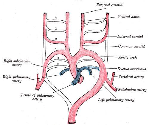
The aortic arches or pharyngeal arch arteries (once referred to as branchial arches in human embryos) are a series of six paired embryological vascular structures which give rise to the great arteries of the neck and head. They are ventral to the dorsal aorta and arise from the aortic sac.
Contents
The aortic arches are formed sequentially within the pharyngeal arches and initially appear symmetrical on both sides of the embryo, but then undergo a significant remodelling to form the final asymmetrical structure of the great arteries.
Arches 1 and 2
The first and second arches disappear early, remnant of the 1st arch forms part of the maxillary artery (branch of external carotid art.) but the dorsal end of the second gives origin to the stapedial artery, a vessel which atrophies in humans but persists in some mammals. It passes through the ring of the stapes and divides into supraorbital, infraorbital, and mandibular branches which follow the three divisions of the trigeminal nerve. The infraorbital and mandibular arise from a common stem, the terminal part of which anastomoses with the external carotid.
On the obliteration of the stapedial artery this anastomosis enlarges and forms the internal maxillary artery, and the branches of the stapedial artery are now branches of this vessel.
The common stem of the infraorbital and mandibular branches passes between the two roots of the auriculotemporal nerve and becomes the middle meningeal artery; the original supraorbital branch of the stapedial is represented by the orbital twigs of the middle meningeal.
Note that the external carotid buds from the horns of the aortic sac left behind by the regression of the first two arches.
Arch 3
The third aortic arch constitutes the commencement of the internal carotid artery, and is therefore named the carotid arch. Common carotid artery and proximal internal carotid artery.
Arch 4
The fourth right arch forms the right subclavian as far as the origin of its internal mammary branch; while the fourth left arch constitutes the arch of the aorta between the origin of the left carotid artery and the termination of the ductus arteriosus.
Arch 5
The fifth arch either never forms or forms incompletely and then regresses.
Arch 6
The proximal part of the sixth right arch persists as the proximal part of the right pulmonary artery while the distal section degenerates; The sixth left arch gives off the left pulmonary artery and forms the ductus arteriosus; this duct remains pervious during the whole of fetal life, but then closes within the first few days after birth due to increased O2 concentration. Oxygen concentration causes the production of bradykinin which causes the ductus to constrict occluding all flow. Within 1–3 months, the ductus is obliterated and becomes the ligamentum arteriosum.
The ductus arteriosus connects at a junction point that has a low pressure zone (commonly called Bernoulli's principle) created by the inferior curvature (inner radius) of the artery. This low pressure region allows the artery to receive (siphon) the blood flow from the pulmonary artery which is under a higher pressure. However, it is extremely likely that the major force driving flow in this artery is the markedly different arterial pressures in the pulmonary and systemic circulations due to the different arteriolar resistances.
His showed that in the early embryo the right and left arches each gives a branch to the lungs, but that later both pulmonary arteries take origin from the left arch.
Great arteries anomalies
Most defects of the great arteries arise as a result of persistence of aortic arches that normally should regress or regression of arches that normally shouldn't.