
 | ||
Right-sided aortic arch is a rare anatomical variant in which the aortic arch is on the right side rather than on the left. During normal embryonic development, the aortic arch is formed by the left fourth aortic arch and the left dorsal aorta. In people with a right-sided aortic arch, instead the right dorsal aorta persists and the distal left aorta disappears.
Contents
Classification
Several types of right-sided aortic arch exist, the most common ones being right-sided aortic arch with aberrant left subclavian artery and the mirror-image type. The variant with aberrant left subclavian artery is associated with congenital heart disease in only a small minority of affected people. The mirror-image type of right aortic arch is very strongly associated with congenital heart disease, in most cases tetralogy of Fallot.
Pathophysiology
The causes of right-sided aortic arch are still unknown. 22q11 deletions have been found in some patients.
Symptoms
A right-sided aortic arch does not cause symptoms on itself, however when it is accompanied by other vascular abnormalities, it may form a vascular ring, causing symptoms due to compression of the trachea and/or esophagus.
Diagnosis
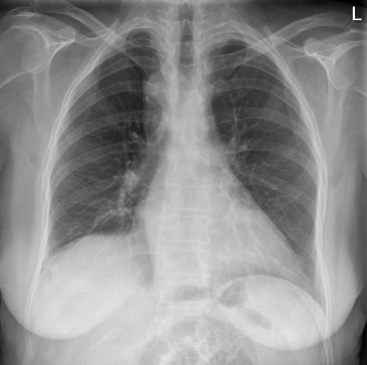
During pregnancy, prenatal ultrasound may reveal the abnormal course of the arch. On chest radiography, a right-sided aortic arch is visualized by the aortic knob (the prominent shadow of the aortic arch) that is located right from the sternum instead of left. Complex lesions are often assessed by MRI or CT.
Epidemiology
Right-sided aortic arch is rare, with a prevalence among adults of about 0.01%.