
Latin Arcus aortae | ||
 | ||
Precursor Fourth left pharyngeal arch artery Branches Brachiocephalic trunkLeft common carotid arteryLeft subclavian arteryContinues as descending aorta, thoracic part | ||
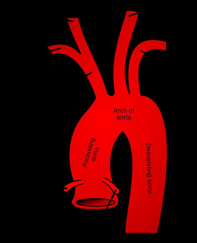
The aortic arch, arch of the aorta or the transverse aortic arch ( /eɪˈɔːrtɪk/) is the part of the aorta between the ascending and descending aorta. The arch travels backward, so that it ultimately runs to the left of the trachea.
Contents
Structure
It begins at the level of the upper border of the second sternocostal articulation of the right side, and runs at first upward, backward, and to the left in front of the trachea; then travels backward on the left side of the trachea and finally passes downward on the left side of the body of the fourth thoracic vertebra. At this point the aortic arch continues as the descending aorta.
The aortic arch has three branches. The first, and largest, branch of the arch of the aorta is the brachiocephalic trunk, which is to the right and slightly anterior to the other two branches and originates behind the manubrium of the sternum. Next, the left common carotid artery originates from the aortic arch to the left of the brachiocephalic trunk, then ascends along the left side of the trachea and through the superior mediastinum. Finally, the left subclavian artery comes off of the aortic arch to the left of the left common carotid artery and ascends, with the left common carotid, through the superior mediastinum and along the left side of the trachea. An anatomical variation is that the left vertebral artery can arise from the aortic arch instead of the left subclavian artery.
The arch of the aorta forms two curvatures: one with its convexity upward, the other with its convexity forward and to the left. Its upper border is usually about 2.5 cm. below the superior border to the manubrium sterni. Blood flows from the upper curvature to the upper regions of the body, located above the heart - namely the arms, neck, and head.
Coming out of the heart, the thoracic aorta has a maximum dimension of 40 mm at the root. By the time it becomes the ascending aorta, the diameter should be < 35–38 mm, and 30 mm at the arch. The diameter of the descending aorta should not exceed 25 mm.
The arch of the Aorta lies within the mediastinum.
Development
The aortic arch is the connection between the ascending and descending aorta, and its central part is formed by the left 4th aortic arch during early development.
The ductus arteriosus connects to the lower part of the arch in foetal life. This allows blood from the right ventricle to mostly bypass the pulmonary vessels as they develop.
The final section of the aortic arch is known as the isthmus of aorta. This is so-called because it is a narrowing (isthmus) of the aorta as a result of decreased blood flow when in foetal life. As the left ventricle of the heart increases in size throughout life, the narrowing eventually dilates to become a normal size. If this does not occur, this can result in coarctation of the aorta. The ductus arteriosus connects to the final section of the arch in foetal life and the ligamentum arteriosum when the ductus arteriosus regresses.
Variation
There are three common variations in how arteries branch from the aortic arch. In about 75% of individuals, the branching is "normal", as described above. In some individuals the left common carotid artery originates from the brachiocephalic artery rather than the aortic arch. In others, the brachiocephalic artery and left common carotid artery share an origin. This variant is found in approximately a 20% of the population. In a third variant, the brachiocephalic artery splits into three arteries: the left common carotid artery, the right common carotid artery and the right subclavian artery; this variant is found in an estimated 7% of individuals.
Clinical significance
The aortic knob is the prominent shadow of the aortic arch on a frontal chest radiograph.
Aortopexy is a surgical procedure in which the aortic arch is fixed to the sternum in order to keep the trachea open.