
Domain Eukarya Higher classification Trichomonas | Genus Trichomonas Rank Species | |
 | ||
Similar Trichomonas, Trichomonadida, Tritrichomonas foetus, Histomonas meleagridis, Tritrichomonas | ||
Suspected frounce trichomonosis trichomonas gallinae trich etc in newfoundland canada
Trichomonas gallinae is a cosmopolitan parasite of pigeons and doves. Other birds such as domestic and wild turkeys, chickens, raptors (hawks, golden eagle, etc.) may also become infected. The disease in pigeons is commonly called canker. The same condition in birds of prey is called frounce. It is believed to be an ancient pathogen causing frounce-like symptoms in dinosaurs.
Contents
- Suspected frounce trichomonosis trichomonas gallinae trich etc in newfoundland canada
- Trichomonas gallinae wmv
- Life cycle
- Transmission
- Pathology
- Clinical signs
- Diagnosis
- Treatment
- Prevention
- References
In 2005, Trichomonas gallinae was first recognized as a cause of disease in British finches, with greenfinch and chaffinch most affected, although a range of garden birds have been found to be susceptible to the parasite. Recent studies have shown that up to a third of adult wood pigeons in Spain may carry the disease.
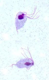
The protozoan has four anterior flagella and an undulating membrane on one side. An important diagnostic feature is the lack of a free posterior flagellum.
Trichomonas gallinae wmv
Life cycle
T. gallinae is generally found in the oral-nasal cavity or anterior end of the digestive and respiratory tracts. The trichomonads multiply rapidly by simple division (binary fission), but do not form a resistant cyst. They therefore die quickly when passed out of the host.
Transmission
Transmission of the parasite from one bird to another occurs in one of three ways:
- Infected parent feeding young
- Contaminated drinking water
- Infected bird is a prey meal for another bird (raptors most commonly)
In pigeons, transmission occurs when infected older birds (carriers) feed "pigeon milk" to newly hatched squabs. Adult birds, which do not show signs of disease, may carry the infection for a year or more and are a constant source of infection for their young.
Turkeys and chickens likely become infected through drinking water or food contaminated with feces, saliva or crop secretions. Because the trichomonads do not survive for long outside the bird, transmission must occur rapidly. Wild pigeons and other birds may be an important source of introducing the infection to domestic birds.
The third method of transmission is more common to birds of prey. An infection may be established in a raptor that has fed on an infected prey bird.
Pathology
Avian trichomoniasis is principally a disease of young birds. T. gallinae varies greatly in its virulence. The severity of the disease depends on the susceptibility of the bird and on the pathogenic potential of the strain of the parasite. Adult birds that recover from the infection may still carry the parasite, but are resistant to reinfection. These birds do not show obvious signs of infection. Interestingly, infection and mortality rates are not closely linked. The disease varies from a mild condition to a rapidly fatal one with death in 4–18 days post infection.
In young birds, the early lesions appear as small white to yellowish areas in the mouth cavity, especially the soft palate. The lesions consist of inflammation and ulceration of the mucosal surface. The lesions increase in size and number and extend to the esophagus, crop and proventriculus. The lesions may develop into large, firm necrotic masses that may block the lumen. Occasionally, the disease may spread by penetrating the underlying tissues to involve the liver and other organs.
The early lesions in the mouth are small, yellowish, circumscribed plaques on the mucosa. More velogenic strains can cause caseated abscessation of the oropharynx. Eventually these space occupying lesions obstruct the esophagus and trachea resulting in emaciation and asphyxiation.
Although lesions are usually seen in the mouth and oropharynx in raptors, it can also affect other mucus membranes. Jessup reports one owl having eye lesions from infection spreading into the nasolacrimal duct. Bony involvement can occur after soft tissue destruction. The organism does not survive posterior to the proventriculus, except in pigeons. Unlike other birds infected with T. gallinae, pigeons are susceptible to secondary organ invasion by virulent strains of the parasite. The visceral form of the disease involves the liver and gastrointestinal tract, causing organ dysfunction.
Clinical signs
In acute cases, there may be little indication that the bird is infected, and death may occur quite suddenly. In other cases, infected pigeon squabs may stop feeding, lose weight, look ruffled and dull, and be unable to stand or maintain their balance. Erosion of the papillae on the palatal flaps is a good sign of infection. Birds may have difficulty swallowing and breathing due to the cheese like deposits in the mouth lining and down the esophagus and trachea.
Diarrhea may also occur. Death may occur within three weeks of infection. Greenish fluid or cheesy material may accumulate in the mouth and crop, and this material may exude from the beak. A pendulous crop may develop in turkey poults and chickens.
Diagnosis
Diagnosis can be accomplished by clinical signs and direct exam or culture. Clinical signs of trichomoniasis include pronounced swallowing motions, excessive salivation and caseous-diphtheritic membranes of the mouth, crop and pharynx. Characteristic yellowish-white nodules in the oral cavity, esophagus and crop strongly suggest trichomoniasis. The infection is confirmed by finding the organism during microscopic examination of the greenish fluids, cheesy material or the lesion. Giemsa staining may aid in identifying the flagellate.
Treatment
2-amino-5-nitrothiazole is currently widely used as it is efficient and there is as yet no resistance.
Antiprotozoal medications, such as dimetridazole and metronidazole, have also been used successfully to treat birds infected with T. gallinae. Treatment usually is successful after 1–2 days. A new culture system called InPouch has practical advantages over the usual in vitro system. If a bird is dyspneic or cannot swallow food, oral plaques can be removed with thumb forceps.
Prevention
Trichomoniasis can be controlled in a flock by culling or treating carrier birds. Food and water sources, as well as bird baths, should be cleaned regularly and protected from contamination by wild pigeons and other birds.
Generally parasitic trichomonads cannot survive drying out. Cleaning feeders and bird baths and leaving them dry for a week may help in decreasing spread of disease although natural infections probably occur in the wider environment. Some related trichomonads do form cysts resistant to drying.
Bird diseases such as trichomoniasis rarely, if ever, affect people; but it is sensible to take care when handling bird feeders by good hand washing after tending the feeders, or wearing suitable disposable gloves. From the sources used, there have been no reports of Trichomonas gallinae infecting humans.