
Specialty rheumatology ICD-9-CM 710.1 DiseasesDB 12845 | ICD-10 M34 OMIM 181750 MedlinePlus 000429 | |
 | ||
Systemic scleroderma, also called diffuse scleroderma or systemic sclerosis, is an autoimmune disease of the connective tissue. It is characterized by thickening of the skin caused by accumulation of collagen, and by injuries to small arteries. There are two forms of scleroderma: localized and systemic. The localized (limited) form affects the skin of only the face, hands, and feet. The systemic (diffuse) form involves those and, in addition, may progress to visceral organs, including the kidneys, heart, lungs, and gastrointestinal tract. Prognosis is determined by the form of the disease and the extent of visceral involvement. Patients with limited cutaneous scleroderma have a 10-year survival rate of 75%; less than 10% develop pulmonary arterial hypertension after 10 to 20 years. Patients with diffuse cutaneous scleroderma have a 10-year survival rate of 55%. Death is most often caused by lung, heart, and kidney involvement. There is also a slight increase in the risk of cancer.
Contents
- Signs and symptoms
- Skin symptoms
- Other organs
- Causes
- Pathophysiology
- Diagnosis
- Treatment
- Topicalsymptomatic
- Kidney disease
- Lung disease and pulmonary hypertension
- Experimental treatments
- Epidemiology
- Patient support groups
- References
Survival rates have greatly increased with effective treatment for kidney failure. Therapies include immunosuppressive drugs and, in some cases, glucocorticoids.
Signs and symptoms
CREST syndrome (calcinosis, Raynaud's phenomenon, esophageal dysfunction, sclerodactyly, and telangiectasia) is associated with limited scleroderma. Other symptoms include:
Skin symptoms
In the skin, systemic sclerosis causes hardening and scarring. The skin may appear tight, reddish, or scaly. Blood vessels may also be more visible. Where large areas are affected, fat and muscle wastage may weaken limbs and affect appearance. Patients report severe and recurrent itching of large skin areas. The severity of these symptoms varies greatly among patients: Some having scleroderma of only a limited area of the skin (such as the fingers) and little involvement of the underlying tissue, while others have progressive skin involvement.
Other organs
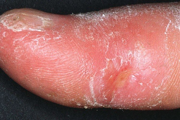
Diffuse scleroderma can cause musculoskeletal, pulmonary, gastrointestinal, renal and other complications. Patients with greater cutaneous involvement are more likely to have involvement of the internal tissues and organs. Most patients (over 80%) have vascular symptoms and Raynaud's phenomenon, which leads to attacks of discoloration of the hands and feet in response to cold. Raynaud's normally affects the fingers and toes. Systemic scleroderma and Raynaud's can cause painful ulcers on the fingers or toes which are known as digital ulcers. Calcinosis (deposition of calcium in lumps under the skin) is also common in systemic scleroderma, and is often seen near the elbows, knees or other joints.
The first joint symptoms that patients with scleroderma have are typically non specific joint pains, which can lead to arthritis, or cause discomfort in tendons or muscles. Joint mobility, especially of the small joints of the hand, may be restricted by calcinosis or skin thickening. Patients may develop muscle weakness, or myopathy, either from the disease or its treatments.
Some impairment in lung function is almost universally seen in patients with diffuse scleroderma on pulmonary function testing; however, it does not necessarily cause symptoms, such as shortness of breath. Some patients can develop pulmonary hypertension, or elevation in the pressures of the pulmonary arteries. This can be progressive, and can lead to right-sided heart failure. The earliest manifestation of this may be a decreased diffusion capacity on pulmonary function testing.
Other pulmonary complications in more advanced disease include aspiration pneumonia, pulmonary hemorrhage and pneumothorax.
Diffuse scleroderma can affect any part of the gastrointestinal tract. The most common manifestation in the esophagus is reflux esophagitis, which may be complicated by peptic stricturing, or benign narrowing of the esophagus. This is best initially treated with proton pump inhibitors for acid suppression, but may require bougie dilatation in the case of stricture.
Scleroderma can decrease motility anywhere in the gastrointestinal tract. The most common source of decreased motility is the esophagus and the lower esophageal sphincter, leading to dysphagia and chest pain. As scleroderma progresses, esophageal involvement from abnormalities in decreased motility may worsen due to progressive fibrosis (scarring). If this is left untreated, acid from the stomach can back up into the esophagus, causing esophagitis and GERD. Further scarring from acid damage to the lower esophagus many times leads to the development of fibrotic narrowing, also known as strictures which can be treated by dilatation, and Barrett's esophagus.
Duodenum: In patients with neuromuscular disorders, particularly progressive systemic sclerosis and visceral myopathy, the duodenum is frequently involved. There may be dilatation, which is often more pronounced in the second, third and fourth parts. The dilated duodenum may be slow to empty and the grossly dilated, atonic organ may produce a sump effect.
The small intestine can also become involved, leading to bacterial overgrowth and malabsorption of bile salts, fats, carbohydrates, proteins, and vitamins. The colon can be involved, and can cause pseudo-obstruction or ischemic colitis.
Rarer complications include pneumatosis cystoides intestinalis, or gas pockets in the bowel wall, wide mouthed diverticula in the colon and esophagus, and liver fibrosis. Patients with severe gastrointestinal involvement can become profoundly malnourished.
Scleroderma may also be associated with gastric antral vascular ectasia (GAVE), also known as watermelon stomach. This is a condition where atypical blood vessels proliferate usually in a radially symmetric pattern around the pylorus of the stomach. GAVE can be a cause of upper gastrointestinal bleeding or iron deficiency anemia in patients with scleroderma.
Renal involvement, in scleroderma, is considered a poor prognostic factor and frequently a cause of death.
The most important clinical complication of scleroderma involving the kidney is scleroderma renal crisis. Symptoms of scleroderma renal crisis are malignant hypertension (high blood pressure with evidence of acute organ damage), hyperreninemia (high renin levels), azotemia (kidney failure with accumulation of waste products in the blood) and microangiopathic hemolytic anemia (destruction of red blood cells). Apart from the high blood pressure, hematuria (blood in the urine) and proteinuria (protein loss in the urine) may be indicative.
In the past scleroderma renal crisis was almost uniformily fatal. While outcomes have improved significantly with the use of ACE inhibitors the prognosis is often guarded, as a significant number of patients are refractory to treatment and develop renal failure. Approximately 5–10% of all diffuse cutaneous scleroderma patients develop renal crisis at some point in the course of their disease. Patients that have rapid skin involvement have the highest risk of renal complications. It is most common in diffuse cutaneous scleroderma, and is often associated with antibodies against RNA polymerase (in 59% of cases). Many proceed to dialysis, although this can be stopped within three years in about a third of cases. Higher age and (paradoxically) a lower blood pressure at presentation make it more likely that dialysis is needed.
Treatments for scleroderma renal crisis include ACE inhibitors. Prophylactic use of ACE inhibitors is currently not recommended, as recent data suggest a poorer prognosis in patient treated with these drugs prior to the development of renal crisis. Transplanted kidneys are known to be affected by scleroderma and patients with early onset renal disease (within one year of the scleroderma diagnosis) are thought to have the highest risk for recurrence.
Causes
There is no clear obvious cause for scleroderma and systemic sclerosis. Genetic predisposition appears to be limited: genetic concordance is small; still, there is often a familial predisposition for autoimmune disease. Polymorphisms in COL1A2 and TGF-β1 may influence severity and development of the disease. There is limited evidence implicating cytomegalovirus (CMV) as the original epitope of the immune reaction, as well as parvovirus B19. Organic solvents and other chemical agents have been linked with scleroderma.
One of the suspected mechanisms behind the autoimmune phenomenon is the existence of microchimerism, i.e. fetal cells circulating in maternal blood, triggering an immune reaction to what is perceived as foreign material.
A distinct form of scleroderma and systemic sclerosis may develop in patients with chronic renal failure. This form, nephrogenic fibrosing dermopathy or nephrogenic systemic fibrosis, has been linked to exposure to gadolinium-containing radiocontrast.
Bleomycin (a chemotherapeutic agent) and possibly taxane chemotherapy may cause scleroderma, and occupational exposure to solvents has been linked with an increased risk of systemic sclerosis.
Pathophysiology
Overproduction of collagen is thought to result from an autoimmune dysfunction, in which the immune system starts to attack the kinetochore of the chromosomes. This would lead to genetic malfunction of nearby genes. T cells accumulate in the skin; these are thought to secrete cytokines and other proteins that stimulate collagen deposition. Stimulation of the fibroblast, in particular, seems to be crucial to the disease process, and studies have converged on the potential factors that produce this effect.
A significant player in the process is transforming growth factor (TGFβ). This protein appears to be overproduced, and the fibroblast (possibly in response to other stimuli) also overexpresses the receptor for this mediator. An intracellular pathway (consisting of SMAD2/SMAD3, SMAD4 and the inhibitor SMAD7) is responsible for the secondary messenger system that induces transcription of the proteins and enzymes responsible for collagen deposition. Sp1 is a transcription factor most closely studied in this context. Apart from TGFβ, connective tissue growth factor (CTGF) has a possible role. Indeed, a common CTGF gene polymorphism is present at an increased rate in systemic sclerosis.
Damage to endothelium is an early abnormality in the development of scleroderma, and this too seems to be due to collagen accumulation by fibroblasts, although direct alterations by cytokines, platelet adhesion and a type II hypersensitivity reaction have similarly been implicated. Increased endothelin and decreased vasodilation has been documented.
Jimenez & Derk describe three theories about the development of scleroderma:
Diagnosis
In 1980, the American College of Rheumatology agreed on diagnostic criteria for scleroderma.
Diagnosis is by clinical suspicion, presence of autoantibodies (specifically anti-centromere and anti-scl70/anti-topoisomerase antibodies) and occasionally by biopsy. Of the antibodies, 90% have a detectable anti-nuclear antibody. Anti-centromere antibody is more common in the limited form (80-90%) than in the diffuse form (10%), and anti-scl70 is more common in the diffuse form (30-40%) and in African American patients (who are more susceptible to the systemic form).
Other conditions may mimic systemic sclerosis by causing hardening of the skin. Diagnostic hints that another disorder is responsible include the absence of Raynaud's phenomenon, a lack of abnormalities in the skin on the hands, a lack of internal organ involvement, and a normal antinuclear antibodies test result.
Treatment
There is no cure for scleroderma, though there is treatment for some of the symptoms, including drugs that soften the skin and reduce inflammation. Some patients may benefit from exposure to heat. Holistic care of patient comprising patient education tailored to patient's education level is useful in view of the complex nature of the disease symptoms and progress.
Topical/symptomatic
Topical treatment for the skin changes of scleroderma do not alter the disease course, but may improve pain and ulceration. A range of NSAIDs (nonsteroidal anti-inflammatory drugs) can be used to ease painful symptoms, such as naproxen. There is limited benefit from steroids such as prednisone. Episodes of Raynaud's phenomenon sometimes respond to nifedipine or other calcium channel blockers; severe digital ulceration may respond to prostacyclin analogue iloprost, and the dual endothelin-receptor antagonist bosentan may be beneficial for Raynaud's phenomenon. The skin tightness may be treated systemically with methotrexate and ciclosporin. and the skin thickness treated with penicillamine.
Kidney disease
Scleroderma renal crisis, the occurrence of acute renal failure and malignant hypertension (very high blood pressure with evidence of organ damage) in people with scleroderma, is effectively treated with drugs from the class of the ACE inhibitors. The benefit of ACE inhibitors extends even to those who have to commence dialysis to treat their kidney disease, and may give sufficient benefit to allow the discontinuation of renal replacement therapy.
Lung disease and pulmonary hypertension
Active alveolitis is often treated with pulses of cyclophosphamide, often together with a small dose of steroids. The benefit of this intervention is modest.
Pulmonary hypertension may be treated with epoprostenol, bosentan and possibly aerolized iloprost.
Experimental treatments
Given the difficulty in treating scleroderma, treatments with a smaller evidence base are often tried to control the disease. These include antithymocyte globulin and mycophenolate mofetil; some reports have reported improvements in the skin symptoms as well as delaying the progress of systemic disease, but neither of them have been subjected to large clinical trials.
Autologous hematopoietic stem cell transplantation (HSCT) is based on the assumption that autoimmune diseases like systemic sclerosis occur when the white blood cells of the immune system attack the body. In this treatment, stem cells from the patient's blood are extracted and stored to preserve them. The patient's white blood cells are destroyed with cyclophosphamide and rabbit antibodies against the white blood cells. Then the stored blood is returned to the patient's bloodstream to reconstitute a healthy blood and immune system which will not attack the body. The results of a phase 3 trial, the Autologous Stem Cell Transplantation International Scleroderma (ASTIS) trial, with 156 patients were published in 2014. HSCT itself has a high treatment mortality, so in the first year, the survival of patients in the treatment group was lower than the placebo group, but at the end of 10 years, the survival in the treatment group was significantly higher. The authors concluded that HSCT could be effective, if limited to patients who were healthy enough to survive HSCT itself. Therefore, HSCT should be given early in the progression of the disease, before it does damage. Patients with heart disease, and patients who smoked cigarettes, were less likely to survive. Another trial, the Stem Cell Transplant vs. Cyclophosphamide (SCOT) trial, is ongoing.
Epidemiology
Systemic scleroderma is a rare disease with an annual incidence of 1 to 2 per 100,000 individuals in the United States. The interval of peak onset starts at age 30 to 35 and ends at age 50 to 55.
In the United States, the prevalence of systemic scleroderma is about 50,000, with different studies giving different estimates, usually ranging between 40,000 and 165,000.
Annual incidence of systemic sclerosis is 19 per million, and prevalence is 19–75 per 100,000, with a female:male ratio of 3:1 (8:1 in mid- to late childbearing years). Incidence is twice as high among African Americans. The Choctaw Native Americans in Oklahoma have the highest prevalence in the world (469 per 100,000).
The disease has some hereditary association. It may also be caused by an immune reaction to a virus (molecular mimicry) or by toxins.
Patient support groups
The Juvenile Scleroderma Network is an organization dedicated to provide emotional support and educational information to parents and their children living with juvenile scleroderma, to support pediatric research to identify the cause of and the cure for juvenile scleroderma, and to enhance public awareness.
In the US, the Scleroderma Research Foundation is dedicated to raise awareness of the disease and assist those who are affected. The Scleroderma Research Foundation sponsors research into the condition. Comedian and television presenter Bob Saget, a board member of the SRF, directed the 1996 ABC TV movie For Hope, starring Dana Delany, which depicts a young woman fatally affected by scleroderma; the film was based on the experiences of Saget's sister Gay.
The Scleroderma Society is a UK charity founded in 1982 to provide support for both people with scleroderma and their families.