
 | ||
Microchimerism (abbreviated Mc) is the presence of a small number of cells that originate from another individual and are therefore genetically distinct from the cells of the host individual. This phenomenon may be related to certain types of autoimmune diseases; however, the mechanisms responsible for this relationship are unclear.
Contents
Human
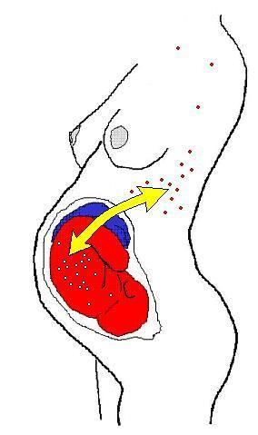
In humans (and perhaps in all placentals) the most common form is fetomaternal microchimerism (also known as fetal cell microchimerism or fetal chimerism) whereby cells from a fetus pass through the placenta and establish cell lineages within the mother. Fetal cells have been documented to persist and multiply in the mother for several decades. The exact phenotype of these cells is unknown, although several different cell types have been identified, such as various immune lineages, mesenchymal stem cells, and placental-derived cells. A 2012 study at the Fred Hutchinson Cancer Research Center, Seattle, has detected cells with the Y chromosome in multiple areas of the brains of deceased women.
Fetomaternal microchimerism occurs during pregnancy and shortly after giving birth for most women. However, not all women who have had children contain fetal cells. Studies suggest that fetomaternal microchimerism could be influenced by killer-cell immunoglobin-like (KIR) ligands. Lymphocytes also influence the development of persisting fetomaternal microchimerism since natural killer cells compose about 70% of lymphocytes in the first trimester of pregnancy. KIR patterns on maternal natural killer cells of the mother and KIR ligands on the fetal cells could have an effect on fetomaternal microchimerism. In one study, mothers with KIR2DS1 exhibited higher levels of fetomaternal microchimerism compared to mothers who were negative for this activating KIR.
The potential health consequences of these cells are unknown. One hypothesis is that these fetal cells might trigger a graft-versus-host reaction leading to autoimmune disease. This offers a potential explanation for why many autoimmune diseases are more prevalent in middle-aged women. Another hypothesis is that fetal cells home to injured or diseased maternal tissue where they act as stem cells and participate in repair. It is also possible that the fetal cells are merely innocent bystanders and have no effect on maternal health.
After giving birth, about 50–75% of women carry fetal immune cell lines. Maternal immune cells are also found in the offspring yielding in maternal→fetal microchimerism, though this phenomenon is about half as frequent as the former.
Microchimerism had also been shown to exist after blood transfusions to a severely immunocompromised population of patients who suffered trauma.
Other possible sources of microchimerism include an individual's older sibling, twin sibling, or vanished twin, with the cells being received in utero. It is hypothesized that unprotected intercourse may be another source of microchimerism, although this has not been shown definitively. Fetal-maternal microchimerism is especially prevalent after abortion or miscarriage.
Animal
Microchimerism occurs in most pairs of twins in cattle. In cattle (and other bovines), the placentae of fraternal twins usually fuse and the twins share blood circulation, resulting in exchange of cell lines. If the twins are a male-female pair, the male hormones from the bull calf have the effect of partially masculinising the heifer (female), creating a martin heifer or freemartin. Freemartins appear female, but are infertile and so cannot be used for breeding or dairy production. Microchimerism provides a method of diagnosing the condition, because male genetic material can be detected in a blood sample.
Relationship with autoimmune diseases and breast cancer
Microchimerism has been implicated in autoimmune diseases. Independent studies repeatedly suggested that microchimeric cells of fetal origin may be involved in the pathogenesis of systemic sclerosis. Moreover, microchimeric cells of maternal origin may be involved in the pathogenesis of a group of autoimmune diseases found in children, i.e. juvenile idiopathic inflammatory myopathies (one example would be juvenile dermatomyositis). Microchimerism has now been further implicated in other autoimmune diseases, including systemic lupus erythematosus. Contrarily, an alternative hypothesis on the role of microchimeric cells in lesions is that they may be facilitating tissue repair of the damaged organ.
Moreover, fetal immune cells have also been frequently found in breast cancer stroma as compared to samples taken from healthy women. It is not clear, however, whether fetal cell lines promote the development of tumors or, contrarily, protect women from developing breast carcinoma.
Systemic lupus erythematosus
The presence of fetal cells in mothers can be associated with benefits when it comes to certain autoimmune diseases. In particular, male fetal cells are related to helping mothers with systemic lupus erythematosus. When kidney biopsies were taken from patients with lupus nephritis, DNA was extracted and run with PCR. The male fetal DNA was quantified and the presence of specific Y chromosome sequences were found. Women with lupus nephritis containing male fetal cells in their kidney biopsies exhibited better renal system functioning. Levels of serum creatinine, which is related to kidney failure, were low in mothers with high levels of male fetal cells. In contrast, women without male fetal cells who had lupus nephritis showed a more serious form of glomerulonephritis and higher levels of serum creatinine.
The specific role that fetal cells play in microchimerism related to certain autoimmune diseases is not fully understood. However, one hypothesis states that these cells supply antigens, causing inflammation and triggering the release of different foreign antigens. This would trigger autoimmune disease instead of serving as a therapeutic. A different hypothesis states that fetal microchimeric cells are involved in repairing tissues. When tissues get inflamed, fetal microchimeric cells go to the damaged site and aid in repair and regeneration of the tissue.
Thyroid disease
Fetal maternal microchimerism may be related to autoimmune thyroid diseases. There have been reports of fetal cells in the lining of the blood and thyroid glands of patients with autoimmune thyroid disease. These cells could become activated after delivery of the baby after immune suppression in the mother is lost, suggesting a role of fetal cells in the pathogenesis of such diseases. Two types of thyroid disease, Hashimoto’s Thyroiditis (HT) and Graves’ Disease (GD) show similarities to graft vs host disease which occurs after hematopoietic stem cell transplants. Fetal cells colonize maternal tissues like the thyroid gland and are able to survive many years postpartum. These fetal microchimeric cells in the thyroid show up in the blood of women affected by thyroid diseases.
Animal models
Fetomaternal microchimerism has been shown in experimental investigations of whether fetal cells can cross the blood brain barrier in mice. The properties of these cells allow them to cross the blood brain barrier and target injured brain tissue. This mechanism is possible because umbilical cord blood cells express some proteins similar to neurons. When these umbilical cord blood cells are injected in rats with brain injury or stroke, they enter the brain and express certain nerve cell markers. Due to this process, fetal cells could enter the brain during pregnancy and become differentiated into neural cells. Fetal microchimerism can occur in the maternal mouse brain, responding to certain cues in the maternal body.
Health implications
Fetal microchimerism could have an implication on maternal health. Isolating cells in cultures can alter the properties of the stem cells, but in pregnancy the effects of fetal stem cells can be investigated without in vitro cultures. Once characterized and isolated, fetal cells that are able to cross the blood brain barrier could impact certain procedures. For example, isolating stem cells can be accomplished through taking them from sources like the umbilical cord. These fetal stem cells can be used in intravenous infusion to repair the brain tissue. Hormonal changes in pregnancy alter neurogenesis, which could create favorable environments for fetal cells to respond to injury.
The true function on fetal cells in mothers is not fully known, however, there have been reports of positive and negative health effects. The sharing of genes between the fetus and mother may lead to benefits. Due to not all genes being shared, health complications may arise as a result of resource allocation. During pregnancy, fetal cells are able to manipulate the maternal system to draw resources from the placenta, while the maternal system tries to limit it.