
NeuroNames hier-565 TA A14.1.05.415 | Dorlands/Elsevier n_11/12582125 FMA 72247 | |
 | ||
Latin nucleus olivaris superior NeuroLex ID Superior olivary complex | ||
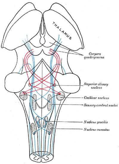
The superior olivary complex (or SOC or superior olive) is a collection of brainstem nuclei that functions in multiple aspects of hearing and is an important component of the ascending and descending auditory pathways of the auditory system. The SOC is intimately related to the trapezoid body: most of the cell groups of the SOC are dorsal (posterior in primates) to this axon bundle while a number of cell groups are embedded in the trapezoid body. Overall, the SOC displays a significant interspecies variation, being largest in bats and rodents and smaller in primates.
Contents
- Physiology
- Relationship to auditory system
- Primary nuclei
- Medial superior olive MSO
- Lateral superior olive LSO
- Medial nucleus of the trapezoid body MNTB
- Periolivary nuclei
- Ventral nucleus of the trapezoid body VNTB
- Lateral nucleus of the trapezoid body LNTB
- Superior periolivary nucleus SPON dorsomedial periolivary nucleus DMPO
- Dorsal periolivary nucleus DPO
- Dorsolateral periolivary nucleus DLPO
- Ventrolateral periolivary nucleus VLPO
- Anterolateral periolivary nucleus ALPO
- Ventromedial periolivary nucleus VMPO
- Rostral periolivary nucleus RPO anterior periolivary nucleus APO
- Caudal periolivary nucleus CPO posterior periolivary nucleus PPO
- Posteroventral periolivary nucleus PVPO
- Pathophysiology
- References
Physiology
The superior olivary nucleus plays a number of roles in hearing. The medial superior olive (MSO) is a specialized nucleus that is believed to measure the time difference of arrival of sounds between the ears (the interaural time difference or ITD). The ITD is a major cue for determining the azimuth of sounds, i.e., localising them on the azimuthal plane – their degree to the left or the right.
The lateral superior olive (LSO) is believed to be involved in measuring the difference in sound intensity between the ears (the interaural level difference or ILD). The ILD is a second major cue in determining the azimuth of high-frequency sounds.
Relationship to auditory system
The superior olivary complex is generally located in the pons, but in humans extends from the rostral medulla to the mid-pons and receives projections predominantly from the anteroventral cochlear nucleus via the trapezoid body, although the posteroventral nucleus projects to the SOC via the intermediate acoustic stria. The SOC is the first major site of convergence of auditory information from the left and right ears.
Primary nuclei
The superior olivary complex is divided into three primary nuclei, the MSO, LSO, and the Medial nucleus of the trapezoid body, and several smaller periolivary nuclei. These three nuclei are the most studied, and therefore best understood. Typically, they are regarded as forming the ascending azimuthal localization pathway.
Medial superior olive (MSO)
The medial superior olive is thought to help locate the azimuth of a sound, that is, the angle to the left or right where the sound source is located. Sound elevation cues are not processed in the olivary complex. The fusiform cells of the dorsal cochlear nucleus (DCN), which are thought to contribute to localization in elevation, bypass the SOC and project directly to the inferior colliculus. Only horizontal data is present, but it does come from two different ear sources, which aids in the localizing of sound on the azimuth axis. The way in which the superior olive does this is by measuring the differences in time between two ear signals recording the same stimulus. Traveling around the head takes about 700 μs, and the medial superior olive is able to distinguish time differences much smaller than this. In fact, it is observed that people can detect interaural differences down to 10 μs. The nucleus is tonotopically organized, but the azimuthal receptive field projection is "most likely a complex, nonlinear map".
The projections of the medial superior olive terminate densely in the ipsilateral central nucleus of the inferior colliculus (ICC). The majority of these axons are considered to be "round shaped" or type R. These R axons are mostly glutamatergic and contain round synaptic vesicles and form asymmetric synaptic junctions.
Lateral superior olive (LSO)
This olive has similar functions to the medial superior olive, but employs intensity to localize the sound source. The LSO receives excitatory, glutamatergic input from spherical bushy cells in the ipsilateral cochlear nucleus and inhibitory, glycinergic input from the medial nucleus of the trapezoid body (MNTB). The MNTB is driven by excitatory input from globular bushy cells in the contralateral cochlear nucleus. Thus, the LSO receives excitatory input from the ipsilateral ear and inhibitory input from the contralateral ear. This is the basis of ILD sensitivity. Projections from both cochlear nuclei are primarily high frequency, and these frequencies are subsequently represented by the majority of LSO neurons (>2/3 over 2–3 kHz in cat). It should be noted that the LSO does in fact encode frequency across the animals audible range (not just "high" frequency). Additional inputs derive from the ipsilateral LNTB (glycinergic, see below), which provide inhibitory information from the ipsilateral cochlear nucleus. Another possibly inhibitory input derives from ipsilateral AVCN non-spherical cells. These cells are either globular bushy or multipolar (stellate). Either of these two inputs could provide the basis for ipsilateral inhibition seen in response maps flanking the primary excitation, sharpening the unit's frequency tuning.
The LSO projects bilaterally to the central nucleus of the inferior colliculus (ICC). Ipsilateral projections are primarily inhibitory (glycinergic), and the contralateral projections are excitatory. Additional projection targets include the dorsal and ventral nuclei of the lateral lemniscus (DNLL & VNLL). The GABAergic projections from the DNLL form a major source of GABA in the auditory brainstem, and project bilaterally to the ICC and to the contralateral DNLL. These converging excitatory and inhibitory connections may act to decrease the level dependence of ILD sensitivity in the ICC compared to the LSO.
Additional projections form the lateral olivocochlear bundle (LOC), which innervates cochlear inner hair cells. These projections are thought to have a long time constant, and act to normalize the sound level detected by each ear in order to aid in sound localization. Considerable species differences exist: LOC projection neurons are distributed within the LSO in rodents, and surround the LSO in predators (i.e. cat).
Medial nucleus of the trapezoid body (MNTB)
Periolivary nuclei
The SOC is composed of between six and nine periolivary nuclei, depending upon the researcher cited, typically named based upon their location with regard to the primary nuclei. These nuclei surround each of the primary nuclei, and contribute to both the ascending and descending auditory systems. These nuclei also form the source of the olivocochlear bundle, which innervates the cochlea. In the guinea pig, ascending projections to the inferior colliculi are primarily ipsilateral (>80%), with the largest single source coming from the SPON. Also, ventral nuclei (RPO, VMPO, AVPO, & VNTB) are almost entirely ipsilateral, while the remaining nuclei project bilaterally.
Ventral nucleus of the trapezoid body (VNTB)
Lateral nucleus of the trapezoid body (LNTB)
Superior periolivary nucleus (SPON) (dorsomedial periolivary nucleus (DMPO))
Dorsal periolivary nucleus (DPO)
Dorsolateral periolivary nucleus (DLPO)
Ventrolateral periolivary nucleus (VLPO)
Anterolateral periolivary nucleus (ALPO)
Ventromedial periolivary nucleus (VMPO)
Rostral periolivary nucleus (RPO) (anterior periolivary nucleus (APO))
Caudal periolivary nucleus (CPO) (posterior periolivary nucleus (PPO))
Posteroventral periolivary nucleus (PVPO)
Pathophysiology
An autopsy of a 21-year-old woman with autism, epilepsy and mental retardation found a near-complete absence of the superior olive.