
Latin vena subclavia TA A12.3.08.002 | MeSH A07.231.908.877 | |
 | ||
Source | ||
The subclavian vein is a paired large vein, one on either side of the body. Their diameter is approximately that of the smallest finger.
Contents
Structure
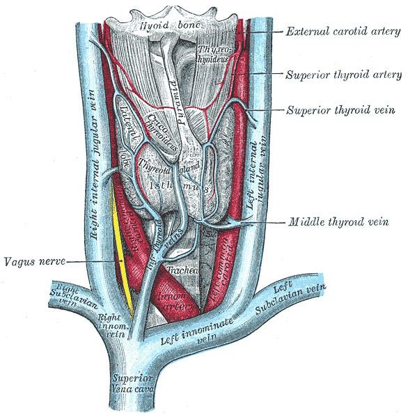
Each subclavian vein is a continuation of the axillary vein and runs from the outer border of the first rib to the medial border of anterior scalene muscle. From here it joins with the internal jugular vein to form the brachiocephalic vein (also known as "innominate vein"). The angle of union is termed the venous angle.
The subclavian vein follows the subclavian artery and is separated from the subclavian artery by the insertion of anterior scalene. Thus, the subclavian vein lies anterior to the anterior scalene while the subclavian artery lies posterior to the anterior scalene (and anterior to the middle scalene).
Function
The thoracic duct drains into the left subclavian vein, near its junction with the left internal jugular vein. It carries lymph (water and solutes) from the lymphatic system, as well as chylomicrons or chyle, formed in the intestines from dietary fat and lipids.
The right lymphatic duct drains its lymph into the junction of the right internal jugular vein, and the right subclavian vein.
History
Sub (below), and clavian (pertaining to the clavicle).