
Source cisterna chyli MeSH Thoracic+Duct TA A12.4.01.007 | Latin ductus thoracicus Dorlands
/Elsevier Thoracic duct | |
 | ||
Drains to junction of the left subclavian vein and left internal jugular vein | ||
In human anatomy, the thoracic duct is the largest lymphatic vessel of the lymphatic system. It is also known as the left lymphatic duct, alimentary duct, chyliferous duct, and Van Hoorne's canal. It carries chyle, a liquid containing both lymph and emulsified fats, rather than pure lymph. Thus when it ruptures, the resulting flood of liquid into the pleural cavity is known as chylothorax.
Contents
In adults, the thoracic duct is typically 38–45 cm in length and has an average diameter of about 5 mm. The vessel usually starts from the level of the twelfth thoracic vertebrae (T12) and extends to the root of the neck. It drains into the systemic (blood) circulation at the angle of the left subclavian and internal jugular veins as a single trunk, at the commencement of the brachiocephalic vein. It also collects most of the lymph in the body other than from the right thorax, arm, head, and neck which are drained by the right lymphatic duct.
Structure
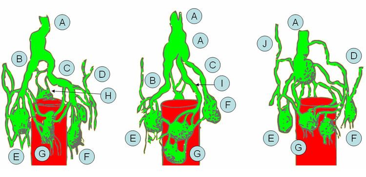
The lymph duct originates in the abdomen from the confluence of the right and left lumbar trunks and the intestinal trunk, forming a significant pathway upward called the cisterna chyli. It traverses the diaphragm at the aortic aperture and ascends the superior and posterior mediastinum between the descending thoracic aorta (to its left) and the azygos vein (to its right). The duct extends vertically in the chest and curves posteriorly to the left carotid artery and left internal jugular vein at the T5 vertebral level It drains into the systemic (blood) circulation at the angle of the left subclavian and internal jugular veins as a single trunk, at the commencement of the brachiocephalic vein, below the clavicle, near the shoulders.
Function
The lymph transport, in the thoracic duct, is mainly caused by the action of breathing, aided by the duct's smooth muscle and by internal valves which prevent the lymph from flowing back down again. There are also two valves at the junction of the duct with the left subclavian vein, to prevent the flow of venous blood into the duct. In adults, the thoracic duct transports up to 4 L of lymph per day.
Clinical significance
The first sign of a malignancy, especially an intraabdominal one, may be an enlarged Virchow's node, a lymph node in the left supraclavicular area, in the vicinity where the thoracic duct empties into the left brachiocephalic vein, right between where the left subclavian vein and left internal jugular join (i.e. the left Pirogoff angle). When the thoracic duct is blocked or damaged a large amount of lymph can quickly accumulate in the pleural cavity, this situation is called chylothorax.