
MeSH A05.360.444.849.789 | ||
 | ||
A Sertoli cell (a kind of sustentacular cell) is a "nurse" cell of the testicles that is part of a seminiferous tubule and helps in the process of spermatogenesis; that is, the production of sperm.
Contents
- Functions
- Secretory
- Structural
- Other functions
- Production of Sertoli cells
- Nomenclature
- Histology
- Pathology
- References
It is activated by follicle-stimulating hormone (FSH) secreted by the adenohypophysis, and has FSH receptor on its membranes. It is specifically located in the convoluted seminiferous tubules (since this is the only place in the testes where the spermatozoa are produced). Development of Sertoli cells is directed by the testis-determining factor protein.
Functions
Because its main function is to nourish the developing sperm cells through the stages of spermatogenesis, the Sertoli cell has also been called the "mother" or "nurse" cell. Sertoli cells also act as phagocytes, consuming the residual cytoplasm during spermatogenesis. Translocation of germ cells from the base to the lumen of the seminiferous tubules occurs by conformational changes in the lateral margins of the Sertoli cells.
Secretory
Sertoli cells secrete the following substances:
Structural
The occluding junctions of Sertoli cells form the blood-testis barrier, a structure that partitions the interstitial blood compartment of the testis from the adluminal compartment of the seminiferous tubules. Because of the apical progression of the spermatogonia (sperm stem cells), the occluding junctions must be dynamically reformed and broken to allow the immunoidentical spermatogonia to cross through the blood-testis barrier so they can become immunologically unique. Sertoli cells control the entry and exit of nutrients, hormones and other chemicals into the tubules of the testis as well as make the adluminal compartment an immune-privileged site.
The cell is also responsible for establishing and maintaining the spermatogonial stem cell niche, which ensures the renewal of stem cells and the differentiation of spermatogonia into mature germ cells that progress stepwise through the long process of spermatogenesis, ending in the release of spermatozoa in a process known as spermiation. Sertoli cells bind to spermatogonial cells via N-cadherins and galctosyltransferase (via carbohydrate residues).
Other functions
During the maturation phase of spermiogenesis, the Sertoli cells consume the unneeded portions of the spermatozoa.
Production of Sertoli cells
Sertoli cells are required for male sexual development. During male development, the gene SRY activates SOX9, which then activates and forms a feedforward loop with FGF9. Sertoli cell proliferation and differentiation is mainly activated by FGF9. The absence of FGF9 tends to cause a female to develop
Once fully differentiated, the Sertoli cell has been considered to be terminally differentiated, and is unable to proliferate. Therefore, once spermatogenesis has begun, no more Sertoli cells are created.
Recently however, some scientists have found a way to induce Sertoli cells to a juvenile proliferative phenotype outside of the body. This gives rise to the possibility of repairing some defects that cause male infertility.
It has been suggested that Sertoli cells may derive from the fetal mesonephros.
Nomenclature
Sertoli cells are called so because of their eponym Enrico Sertoli, an Italian physiologist who discovered them while studying medicine in the University of Pavia, Italy.
He published a description of this cell in 1865. The cell was discovered by Sertoli with a Belthle microscope purchased in 1862, which he used while studying medicine.
In the 1865 publication, his first description used the terms "tree-like cell" or "stringy cell" and most importantly he referred to these as "mother cells." It was other scientists who used Enrico's family name, Sertoli, to label these cell in publications, starting in 1888. As of 2006, two textbooks that are devoted specifically to the Sertoli cell have been published.
Histology
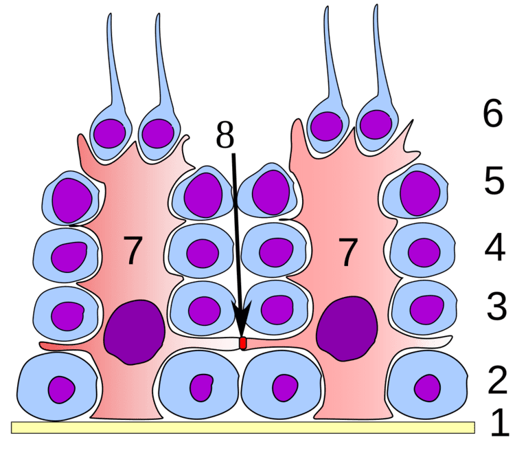
On slides, using standard staining, Sertoli cells can easily be confused with the other cells of the germinal epithelium. The most distinctive feature of the Sertoli cell is the dark nucleolus.
Pathology
Sertoli-Leydig cell tumour is part of the sex cord-stromal tumour group of ovarian neoplasms.