
ICD-9-CM 702.1 DiseasesDB 29386 | ICD-10 L82 OMIM 182000 MedlinePlus 000884 | |
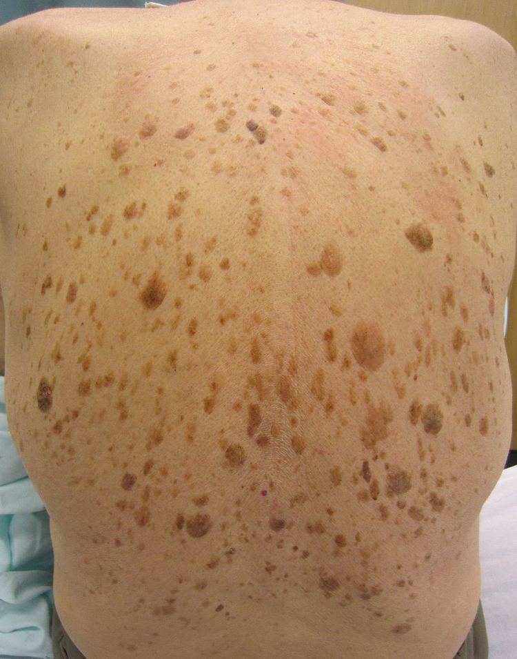 | ||
A seborrheic keratosis, also known as seborrheic verruca or a senile wart, is a non-cancerous (benign) skin tumor that originates from cells in the outer layer of the skin (keratinocytes). Like liver spots, seborrheic keratoses are seen more often as people age.
Contents
The tumors (also called lesions) appear in various colors, from light tan to black. They are round or oval, feel flat or slightly elevated, like the scab from a healing wound, and range in size from very small to more than 2.5 centimetres (1 in) across. They can often come in association with other skin conditions, including basal cell carcinoma. Sometimes seborrheic keratosis and basal cell carcinoma occur at the same location, and sometimes seborrheic keratosis progresses to basal cell carcinoma. At clinical examination the differential diagnosis include warts and melanoma. Because only the top layers of the epidermis are involved, seborrheic keratoses are often described as having a "pasted on" appearance. Some dermatologists refer to seborrheic keratoses as "seborrheic warts", because they resemble warts, but strictly speaking the term "warts" refers to lesions that are caused by human papillomavirus.
Classification
Seborrheic keratoses may be divided into the following types:
Also see:
Cause
The cause of seborrheic keratosis is not known.
Diagnosis
Visual diagnosis is made by the "stuck on" appearance, horny pearls or cysts embedded in the structure. Darkly pigmented lesions can be challenging to distinguish from nodular melanomas. Furthermore, thin seborrheic keratoses on facial skin can be very difficult to differentiate from lentigo maligna even with dermatoscopy. Clinically, epidermal nevi are similar to seborrheic keratoses in appearance. Epidermal nevi are usually present at or near birth. Condylomas and warts can clinically resemble seborrheic keratoses, and dermatoscopy can be helpful. On the penis and genital skin, condylomas and seborrheic keratoses can be difficult to differentiate, even on biopsy.
To date, the gold standard in the diagnosis of seborrheic keratosis is represented by the histolopathologic analysis of a skin biopsy.
Treatment
No treatment of seborrheic keratoses is necessary, except for aesthetic reasons. Since a slightly increased risk of localized infection caused by picking at the lesion has been described, if a lesion becomes itchy or irritated by clothing or jewelry, a surgical excision is generally recommended.
Small lesions can be treated with light electrocautery. Larger lesions can be treated with electrodesiccation and curettage, shave excision, or cryosurgery. When correctly performed, removal of seborrheic keratoses will not cause much visible scarring except in persons with dark skin tones.
Epidemiology
Seborrheic keratosis is the most common benign skin tumor. Incidence increases with age. There is less prevalence in people with darker skin. In large-cohort studies, 100% of the patients over age 50 had at least one seborrhoeic keratosis. Onset is usually in middle age, although they are common in younger patients too—found in 12% of 15-year-olds to 25-year-olds—making the term "senile keratosis" a misnomer.