
Specialty infectious disease ICD-9-CM 066.3 MeSH D012295 | ICD-10 A92.4 DiseasesDB 31094 | |
 | ||
Rift Valley fever (RVF) is a viral disease that can cause mild to severe symptoms. The mild symptoms may include: fever, muscle pains, and headaches which often last for up to a week. The severe symptoms may include: loss of sight beginning three weeks after the infection, infections of the brain causing severe headaches and confusion, and bleeding together with liver problems which may occur within the first few days. Those who have bleeding have a chance of death as high as 50%.
Contents
- Signs and symptoms
- Virology
- Transmission
- Pathogenesis
- Diagnosis
- Prevention
- Epidemiology
- Outbreaks
- 200607 outbreak in Kenya and Somalia
- 2010 South Africa outbreak
- Biological weapon
- Research
- References
The disease is caused by the RVF virus, which is of the Phlebovirus type. It is spread by either touching infected animal blood, breathing in the air around an infected animal being butchered, drinking raw milk from an infected animal, or the bite of infected mosquitoes. Animals such as cows, sheep, goats, and camels may be affected. In these animals it is spread mostly by mosquitoes. It does not appear that one person can infect another person. The disease is diagnosed by finding antibodies against the virus or the virus itself in the blood.
Prevention of the disease in humans is accomplished by vaccinating animals against the disease. This must be done before an outbreak occurs because if it is done during an outbreak it may worsen the situation. Stopping the movement of animals during an outbreak may also be useful, as may decreasing mosquito numbers and avoiding their bites. There is a human vaccine; however, as of 2010 it is not widely available. There is no specific treatment and medical efforts are supportive.
Outbreaks of the disease have only occurred in Africa and Arabia. Outbreaks usually occur during periods of increased rain which increase the number of mosquitoes. The disease was first reported among livestock in Rift Valley of Kenya in the early 1900s, and the virus was first isolated in 1931.
Signs and symptoms
In humans, the virus can cause several syndromes. Usually, sufferers have either no symptoms or only a mild illness with fever, headache, muscle pains, and liver abnormalities. In a small percentage of cases (< 2%), the illness can progress to hemorrhagic fever syndrome, meningoencephalitis (inflammation of the brain and tissues lining the brain), or affect the eye. Patients who become ill usually experience fever, generalised weakness, back pain, dizziness, and weight loss at the onset of the illness. Typically, people recover within two to seven days after onset.
About 1% of people with the disease die of it. In livestock, the fatality level is significantly higher. Pregnant livestock infected with RVF abort virtually 100% of foetuses. An epizootic (animal disease epidemic) of RVF is usually first indicated by a wave of unexplained abortions.
Other signs in livestock include vomiting and diarrhoea, respiratory disease, fever, lethargy, anorexia and sudden death in young animals.
Virology
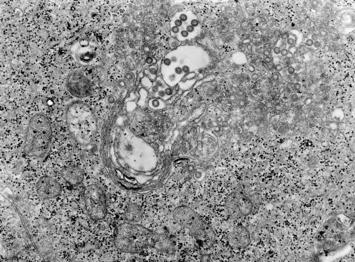
The virus belongs to the Bunyaviridae family. This is a family of enveloped negative single stranded RNA viruses. All Bunyaviruses have an outer lipid envelope with two glycoproteins—G(N) and G(C)—required for cell entry. They deliver their genome into the host-cell cytoplasm by fusing their envelope with an endosomal membrane.
The virus' G(C) protein has a class II membrane fusion protein architecture similar to that found in flaviviruses and alphaviruses. This structural similarity suggests that there may be a common origin for these viral families.
The virus' 11.5 kb tripartite genome is composed of single-stranded RNA. As a Phlebovirus, it has an ambisense genome. Its L and M segments are negative-sense, but its S segment is ambisense. These three genome segments code for six major proteins: L protein (viral polymerase), the two glycoproteins G(N) and G(C), the nucleocapsid N protein, and the nonstructural NSs and NSm proteins.
Transmission
The virus is transmitted through mosquito vectors, as well as through contact with the tissue of infected animals. Two species—Culex tritaeniorhynchus and Aedes vexans—are known to transmit the virus. Other potential vectors include Aedes caspius, Aedes mcintosh, Aedes ochraceus, Culex pipiens, Culex antennatus, Culex perexiguus, Culex zombaensis and Culex quinquefasciatus. Contact with infected tissue is considered to be the main source of human infections. The virus has been isolated from two bat species: the Peter's epauletted fruit bat (Micropteropus pusillus) and the aba roundleaf bat (Hipposideros abae), which are believed to be reservoirs for the virus.
Pathogenesis
Although many components of the RVFV’s RNA play an important role in the virus’ pathology, the nonstructural protein encoded on the S segment (NSs) is the only component that has been found to directly affect the host. NSs is hostile and combative against the hosts interferon (IFNs) antiviral response. IFNs are essential in order for the immune system to fight off viral infections in a host. This inhibitory mechanism is believed to be due to a number of reasons, the first being, competitive inhibition of the formation of the transcription factor. On this transcription factor, NSs interacts with and binds to a subunit that is needed for RNA polymerase I and II. This interaction cause competitive inhibition with another transcription factor component and prevents the assembly process of the transcription factor complex, which results in the suppression of the host antiviral response. Transcription suppression is believed to be another mechanism of this inhibitory process. This occurs when an area of NSs interacts with and binds to the host’s protein, SAP30 and forms a complex. This complex causes histone acetylation to regress, which is needed for transcriptional activation of the IFN promoter. This causes IFN expression to be obstructed. Lastly, NSs has also been known to affect regular activity of double-stranded RNA-dependent protein kinase R.. This protein is involved in cellular antiviral responses in the host. When RVFV is able to enter the hosts DNA, NSs forms a filamentous structure in the nucleus. This allows the virus to interact with specific areas of the hosts DNA that relates to segregation defects and induction of chromosome continuity. This increases host infectivity and decreases the host’s antiviral response.
Diagnosis
Diagnosis relies on viral isolation from tissues, or serological testing with an ELISA. Other methods of diagnosis include Nucleic Acid Testing (NAT), cell culture, and IgM antibody assays. As of September 2016, the Kenya Medical Research Institute (KEMRI) has developed a product called Immunoline, designed to diagnose the disease in humans much faster than in previous methods.
Prevention
A vaccine has been conditionally approved for use in animals in the US. It has been shown that knockout of the NSs and NSm nonstructural proteins of this virus produces an effective vaccine in sheep as well.
Epidemiology
RVF outbreaks occur across sub-Saharan Africa, with outbreaks occurring elsewhere infrequently. In Egypt in 1977–78, an estimated 200,000 people were infected and there were at least 594 deaths. In Kenya in 1998, the virus killed over 400 Kenyans. In September 2000, an outbreak was confirmed in Saudi Arabia and Yemen. On 19 October 2011, a case of Rift Valley fever contracted in Zimbabwe was reported in a Caucasian female traveler who returned to France after a 26-day stay in Marondera, Mashonaland East Province during July and August, 2011 but later classified as "not confirmed."
Outbreaks
Outbreaks of this disease usually correspond with the warm phases of the EI Niño/Southern Oscillation. During this time there is an increase in rainfall, flooding and greenness of vegetation index. This leads to an increase in mosquito vectors and is seen for a number of reasons. RVFV can be transmitted vertically in mosquitos, meaning that the virus can be passed from the mother to her offspring. During dry conditions, the virus is able to remain viable for a number of years in the egg. Mosquitos lay their eggs in water, where they eventually hatch. Since water is essential for mosquito eggs to hatch, it can be understood why rainfall and flooding cause an increase in the mosquito population, and in turn, an increased potential for the virus.
2006/07 outbreak in Kenya and Somalia
In November 2006, a Rift Valley fever outbreak occurred in Kenya. The victims are from the North Eastern Province and Coast Province of Kenya, which had received heavy rain in recent months, causing floods and creating breeding grounds for mosquitoes, which spread the virus of the fever from infected livestock to humans.
By 7 January 2007, about 75 people had died and another 183 were infected. The outbreak forced the closure of livestock markets in the North Eastern Province, affecting the economy of the region.
The outbreak was subsequently reported to have moved into Maragua and Kirinyaga districts of Central Province of Kenya.
On 20 January 2007, the outbreak was reported to have crossed into Somalia from Kenya and killed 14 people in the Lower Jubba region.
As of 23 January 2007, cases had started to crop up at the Kenyan capital, Nairobi. Businesses were suffering large losses, as customers were shunning the common meat joints for the popular nyama choma (roast meat), as it was believed to be spreading the fever.
In December 2006 and again in January 2007, Taiwan International Health Action (Taiwan IHA) began operating missions in Kenya consisting of medical experts assisting in training laboratory and health facility personnel, and included donations of supplies, such as mosquito sprays. The United States Centers for Disease Control also set up an assistance mission and laboratory in Kenya.
By the end of January, 2007, some 148 people had died since the outbreak began in December.
As at 14 March 2007, the Kenyan government declared RVF as having diminished drastically after spending an estimated 2.5 million in vaccine and deployment costs. It also lifted the ban on cattle movement in the affected areas.
As of 2 November 2007, 125 cases, including 60 deaths, had been reported from more than 10 localities of White Nile, Sinnar, and Gezira states in Sudan. Young adult males are predominantly affected. More than 25 human samples have been found positive for RVF by PCR or ELISA.
2010 South Africa outbreak
As of 8 April 2010, the Ministry of Health South Africa had reported 87 human cases infected with Rift Valley fever (RVF), including two deaths in Free State, Eastern Cape and Northern Cape provinces. Most of these cases reported direct contact with RVFV-infected livestock and or were linked to farms with confirmed animal cases of RVF. The human cases are: farmers, veterinarians and farm workers. All cases were confirmed with RVF by test conducted at the National Institute of Communicable Diseases (NICD) in Johannesburg, South Africa.
An ongoing outbreak of Rift Valley fever virus (RVFV) infection is affecting sheep, goats, cattle and wildlife on farms within Free State, Eastern Cape, Northern Cape, Western Cape, Mpumalanga, North West, and Gauteng provinces. As of 29 March 2010, about 78 farms reported laboratory-confirmed animal cases, with extensive livestock deaths.
Outbreak investigations by the Department of Health and the Department of Agriculture, Forestry and Fisheries are ongoing, and are being supported by the South African Field Epidemiology and Training Programme and NICD. The Department of Health and the Department of Agriculture are taking measures to enhance disease surveillance among cattle and in managing the control of the disease outbreak.
Sporadic cases of RVFV infection in animals have been documented in South Africa in recent years. The last major outbreak of the disease in humans occurred between 1974 and 1976, where an estimated 10,000 to 20,000 cases were recorded.
The disease claimed the life of Springbok rugby player Juan Smith's father Giel.
Biological weapon
Rift Valley fever was one of more than a dozen agents that the United States researched as potential biological weapons before the nation suspended its biological weapons program in 1969 due to possible bomb threats that might cause a panic in the United States.
Research
The disease is one of several identified by WHO as a likely cause of a future epidemic in a new plan developed after the Ebola epidemic for urgent research and development toward new diagnostic tests, vaccines and medicines.