
 | ||
The Raman microscope is a laser-based microscopic device used to perform Raman spectroscopy. The term MOLE (molecular optics laser examiner) is used to refer to the Raman-based microprobe. The technique used is named after C. V. Raman who discovered the scattering properties in liquids.
Contents
Configuration
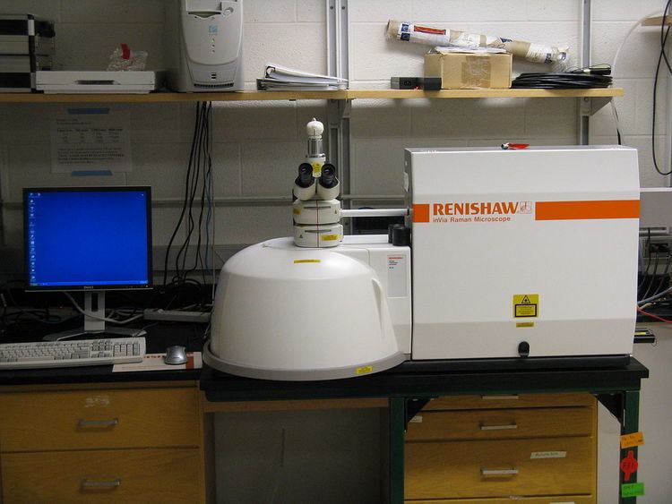
The Raman microscope begins with a standard optical microscope, and adds an excitation laser, laser rejection filters, a spectrometer or monochromator, and an optical sensitive detector such as a charge-coupled device (CCD), or photomultiplier tube, (PMT). Traditionally Raman microscopy was used to measure the Raman spectrum of a point on a sample, more recently the technique has been extended to implement Raman spectroscopy for direct chemical imaging over the whole field of view on a 3D sample.
Imaging modes
In direct imaging, the whole field of view is examined for scattering over a small range of wavenumbers (Raman shifts). For instance, a wavenumber characteristic for cholesterol could be used to record the distribution of cholesterol within a cell culture. The other approach is hyperspectral imaging or chemical imaging, in which thousands of Raman spectra are acquired from all over the field of view. The data can then be used to generate images showing the location and amount of different components. Taking the cell culture example, a hyperspectral image could show the distribution of cholesterol, as well as proteins, nucleic acids, and fatty acids. Sophisticated signal- and image-processing techniques can be used to ignore the presence of water, culture media, buffers, and other interference.
Resolution
Raman microscopy, and in particular confocal microscopy, has very high spatial resolution. For example, the lateral and depth resolutions were 250 nm and 1.7 µm, respectively, using a confocal Raman microspectrometer with the 632.8 nm line from a helium–neon laser with a pinhole of 100 µm diameter. Since the objective lenses of microscopes focus the laser beam to several micrometers in diameter, the resulting photon flux is much higher than achieved in conventional Raman setups. This has the added benefit of enhanced fluorescence quenching. However, the high photon flux can also cause sample degradation, and for this reason some setups require a thermally conducting substrate (which acts as a heat sink) in order to mitigate this process.
Raman imaging
Another tool that is becoming more popular is global Raman imaging. This technique is being used for the characterization of large scale devices, mapping of different compounds and dynamics study. It has already been used for the characterization of graphene layers, J-aggregated dyes inside carbon nanotubes and multiple other 2D materials such as MoS2 and WSe2. Since the excitation beam is dispersed over the whole field of view, those measurements can be done without damaging the sample. By using Raman microspectroscopy, in vivotime- and space-resolved Raman spectra of microscopic regions of samples can be measured. As a result, the fluorescence of water, media, and buffers can be removed. Consequently, in vivotime- and space-resolved Raman spectroscopy is suitable to examine proteins, cells and organs.
Raman microscopy for biological and medical specimens generally uses near-infrared (NIR) lasers (785 nm diodes and 1064 nm Nd:YAG are especially common). This reduces the risk of damaging the specimen by applying higher energy wavelengths. However, the intensity of NIR Raman is low (owing to the ω4 dependence of Raman scattering intensity), and most detectors require very long collection times. Recently, more sensitive detectors have become available, making the technique better suited to general use. Raman microscopy of inorganic specimens, such as rocks and ceramics and polymers, can use a broader range of excitation wavelengths
Correlative Raman imaging
Correlative imaging of any kind is becoming more popular due to enabling a user to get more info from one area of interest. There exist few techniques, which are combined with Raman spectroscopy - AFM, SEM etc.
Correlative SEM-Raman imaging is the integration of a confocal Raman microscope into an SEM chamber which allows correlative imaging of several techniques, such as SE, BSE, EDX, EBSD, EBIC, CL, AFM. Moreover, adding a focused ion beam (FIB) on the chamber allows removal of the material and therefore 3D imaging of the sample. Low-vacuum mode allows analysis on biological and non-conductive samples.