Trisha Shetty (Editor)

Radial nerve

Updated on
Edit
Like
Comment
Share on FacebookTweet on TwitterShare on LinkedInShare on Reddit
From
  
posterior cord

Dorlands /Elsevier
  
n_05/12566581

Latin
  
nervus radialis

Radial nerve

To
  
posterior interosseous nerve

Innervates
  
posterior compartment of the arm, posterior compartment of the forearm

MeSH
  
A08.800.800.720.050.700

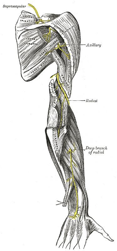
The radial nerve is a nerve in the human body that supplies the posterior portion of the upper limb. It innervates the medial and lateral heads of the triceps brachii muscle of the arm, as well as all 12 muscles in the posterior osteofascial compartment of the forearm and the associated joints and overlying skin.

Contents

It originates from the brachial plexus, carrying fibers from the ventral roots of spinal nerves C5, C6, C7, C8 & T1.

The radial nerve and its branches provide motor innervation to the dorsal arm muscles (the triceps brachii and the anconeus) and the extrinsic extensors of the wrists and hands; it also provides cutaneous sensory innervation to most of the back of the hand, except for the back of the little finger and adjacent half of the ring finger (which are innervated by the ulnar nerve)

The radial nerve divides into a deep branch, which becomes the posterior interosseous nerve, and a superficial branch, which goes on to innervate the dorsum (back) of the hand.

Structure

The radial nerve originates as a terminal branch of the posterior cord of the brachial plexus. It goes through the arm, first in the posterior compartment of the arm, and later in the anterior compartment of the arm, and continues in the posterior compartment of the forearm.

In arm

From the brachial plexus, it travels posteriorly through what is often called the triangular interval (US), the lower triangular space of the axilla (UK) or the triceps hiatus.

Having passed through this inter muscular gap, the radial nerve continues posteriorly in a medial to lateral fashion on the arm while in conjunction with the deep artery of arm.

The nerve will first give off branches to the medial head of the triceps brachii and then enter a groove on the humerus, the radial sulcus (AKA spiral groove), where it innervates the lateral head of the triceps. It is commonly believed that the radial nerve also provided motor innervation to the long head of the triceps. However, a study conducted in 2004 determined that, in 20 cadaveric specimens and 15 surgical dissections on participants, the long head was innervated by a branch of the axillary nerve in all cases.

With the lateral and medial heads of the triceps innervated, the radial nerve emerges from the radial groove on the lateral aspect of the humerus.

At this point, it pierces the lateral intermuscular septum and enters the anterior compartment of the arm.

It then courses inferiorly between the brachialis and brachioradialis muscles.

When the radial nerve reaches the distal part of the humerus, it passes anteriorly to the lateral epicondyle and continues in the forearm.

In forearm

In the forearm, it branches into a superficial branch (primarily sensory) and a deep branch (primarily motor).

  • The superficial branch of the radial nerve descends in the forearm under the brachioradialis. It crosses brachioradialis to enter posterior of forearm near the back of the wrist and supply dorsum of hand. It gives nerve supply to dorsal aspect of thumb, index finger,middle finger and radial side of ring finger except the nail beds, which are supplied by proper digital branches of median nerve.
  • The deep branch of the radial nerve pierces the supinator muscle, winds around the radius under the cover of supinator to reach posterior of forearm where it again pierces supinator and after which it is known as the posterior interosseous nerve of forearm.It pierces the posterior extensor muscles and comes to lie on posterior interosseous membrane just below extensor pollicis brevis. It continues to move along with posterior interosseous artery(a deep branch of common interosseous artery which is a branch of ulnar artery), and ends as a pseudoganglion below extensor retinaculum.
  • Function

    The following are branches of the radial nerve (including the superficial branch of the radial nerve and the deep branch of the radial nerve/posterior interosseous nerve).

    Cutaneous

    Cutaneous innervation by the radial nerve is provided by the following nerve branches:

  • Posterior cutaneous nerve of arm (originates in axilla)
  • Inferior lateral cutaneous nerve of arm (originates in arm)
  • Posterior cutaneous nerve of forearm (originates in arm)
  • The superficial branch of the radial nerve provides sensory innervation to much of the back of the hand, including the web of skin between the thumb and index finger.

    Motor

    Muscular branches of the radial nerve:

  • Triceps brachii (lateral and medial heads)
  • Anconeus
  • Brachioradialis
  • Extensor carpi radialis longus
  • Deep branch of the radial nerve:

  • Extensor carpi radialis brevis
  • Supinator
  • Posterior interosseous nerve (a continuation of the deep branch after the supinator):

  • Extensor digitorum
  • Extensor digiti minimi
  • Extensor carpi ulnaris
  • Abductor pollicis longus
  • Extensor pollicis brevis
  • Extensor pollicis longus
  • Extensor indicis
  • The radial nerve (and its deep branch) provides motor innervation to the muscles in the posterior compartment of the arm and forearm, which are mostly extensors.

    Injury

    Injury to the radial nerve at different levels causes different syndromes with varying motor and sensory deficits.

    At the axilla

  • Common mechanisms of injury: Saturday night palsy, crutch palsy
  • Motor deficit:
  • Loss of extension of forearm, weakness of supination, and loss of extension of hand and fingers.
  • Presence of wrist drop, due to inability to extend the hand and fingers.
  • Sensory deficit: Loss of sensation in lateral arm, posterior forearm, the radial half of dorsum of hand, and dorsal aspect of radial 3 12 digits, excluding their nail beds.
  • At mid-arm

  • Common mechanism of injury: Mid-shaft humeral fracture
  • Motor deficit:
  • Weakness of supination, and loss of extension of hand and fingers.
  • Presence of wrist drop, due to inability to extend the hand and fingers.
  • Sensory deficit: Loss of sensation in posterior forearm, the radial half of dorsum of hand, and dorsal aspect of radial 3 12 digits, excluding their nail beds.
  • Just below the elbow

  • Common mechanism of injury: Neck of radius fracture, elbow dislocation or fracture, tight cast, rheumatoid nodules, injections due to tennis elbow, injuring the deep branch of the radial nerve that pierces the radial head, causing posterior interosseous nerve syndrome
  • Motor deficit:
  • Weakness in extension of hand and loss of extension of fingers.
  • Presence of finger drop, and partial wrist drop, since the extensor carpi radialis longus and brachioradialis muscles are working.
  • Sensory deficit: None, as sensation is supplied by the superficial radial nerve
  • Within the distal forearm:

  • Common mechanism of injury: Wartenberg's syndrome, (not to be confused with Wartenberg's sign), due to nerve entrapment beneath the tendinous insertion of brachioradialis, tight jewellery, and watch bands.
  • Motor deficit: None
  • Sensory deficit: Numbness and tingling in radial half of dorsum of hand, and dorsal aspect of radial 3 12 digits, excluding their nail beds.
  • In Wartenberg's syndrome, there is significant radial wrist pain, and close resemblance to symptoms in de Quervain's tenosynovitis. Finkelstein's test may be positive.
  • References

    Radial nerve Wikipedia