Trisha Shetty (Editor)

Median nerve

Updated on
Edit
Like
Comment
Share on FacebookTweet on TwitterShare on LinkedInShare on Reddit
Latin
  
Nervus medianus

Dorlands /Elsevier
  
n_05/12566162

MeSH
  
Median+Nerve

TA
  
A14.2.03.031

Median nerve

From
  
Lateral cord and Medial cord

Innervates
  
Anterior compartment of the forearm (with two exceptions), Thenar eminence, Lumbricals, skin of the hand

The median nerve is a nerve in humans and other animals in the upper limb. It is one of the five main nerves originating from the brachial plexus.

Contents

The median nerve originates from the lateral and medial cords of the brachial plexus, and has contributions from ventral roots of C5-C7(lateral cord) and C8 & T1 (medial cord).

The median nerve is the only nerve that passes through the carpal tunnel. Carpal tunnel syndrome is the disability that results from the median nerve being pressed in the carpal tunnel.

Structure

After receiving inputs from both the lateral and medial cords of the brachial plexus, the median nerve enters the arm from the axilla at the inferior margin of the teres major muscle. It then passes vertically down and courses with the brachial artery on the medial side of the arm between biceps brachii and brachialis. At first it is lateral to the artery and lies anterior to the elbow joint; it then crosses anteriorly to run medial to the artery in the distal arm and into the cubital fossa.

Inside the cubital fossa the median nerve passes medial to the brachial artery, in front of the point of insertion of the brachialis muscle and deep to the biceps.

The median nerve gives off an articular branch in the upper arm as it passes the elbow joint. A branch to pronator teres may arise from the median nerve immediately proximal to the elbow joint.

Forearm

The median nerve arises from the cubital fossa and passes between the two heads of pronator teres. It then travels between flexor digitorum superficialis and flexor digitorum profundus before emerging between flexor digitorum superficialis and flexor pollicis longus.

The unbranched portion of the median nerve (which arises from the cubital fossa) innervates muscles of superficial and intermediate groups of the anterior(flexor) compartment except flexor carpi ulnaris.

The median nerve does give off two branches as it courses through the forearm:

  • The anterior interosseous branch courses with the anterior interosseous artery and innervates all the muscles of the deep group of the anterior compartment of the forearm except the medial (ulnar half,which is supplied by ulnar nerve) half of flexor digitorum profundus and flexor carpi ulnaris muscle. It ends with its innervation of pronator quadratus.
  • The palmar cutaneous branch of the median nerve arises at the distal part of the forearm. It supplies sensory innervation to the lateral aspect of the skin of the palm (but not the digits).
  • Hand

    The median nerve enters the hand through the carpal tunnel, deep to the flexor retinaculum along with the tendons of flexor digitorum superficialis, flexor digitorum profundus, and flexor pollicis longus.

    From there it sends off several branches:

  • 1. Recurrent branch to muscles of the thenar compartment (the recurrent branch is also called "the million dollar nerve"). Here it provides motor innervation to opponens pollicis, abductor pollicis brevis and superficial part of flexor pollicis brevis.
  • 2. Digital cutaneous branches to common palmar digital branch and proper palmar digital branch of the median nerve which supply the:
  • a) lateral (radial) three and a half digits on the palmar side
  • b) dorsum of the tips of index, middle and thumb
  • 3. The median nerve supplies motor innervation to the first and second Lumbricals of the hand.
  • Variation

    There are multiple naturally occurring anomalies of the median nerve.

  • Bifurcation of the median nerve typically occurs after the nerve exits the carpal tunnel; however, in a small percentage (5%-10%) of individuals, the median nerve bifurcates more proximal in the carpal tunnel, wrist, or forearm.
  • During gestation, a median artery that serves the hand retracts. However, in some individuals the median artery does not retract and follows the course next to the median nerve into the hand.
  • Martin-Gruber anastomoses can occur when branches of the median nerve cross-over in the forearm and merge with the ulnar nerve to innervate portions of the forehand.
  • Riche-Cannieu anastomoses can occur when there is connection between recurrent branch of the median nerve and deep branch of the ulnar nerve of the hand.
  • Arm

    The median nerve has no voluntary motor or cutaneous function in the brachium. It gives vascular branches to the wall of the brachial artery. These vascular branches carry sympathetic fibers.

    Forearm

    It innervates all of the flexors in the forearm except flexor carpi ulnaris and that part of flexor digitorum profundus that supplies the 4th and 5th digits. The latter two muscles are supplied by the ulnar nerve (specifically the Muscular branches of ulnar nerve).

    The main portion of the median nerve supplies the following muscles:

    Superficial group:

  • Pronator teres
  • Flexor carpi radialis
  • Palmaris longus
  • Intermediate group:

  • Flexor digitorum superficialis muscle
  • The anterior interosseus branch of the median nerve supplies the following muscles:

    Deep group:

  • Flexor digitorum profundus (only the lateral half)
  • Flexor pollicis longus
  • Pronator quadratus
  • Hand

    In the hand, the median nerve supplies motor innervation to the 1st and 2nd lumbrical muscles. It also supplies the muscles of the thenar eminence by a recurrent thenar branch. The rest of the intrinsic muscles of the hand are supplied by the ulnar nerve.

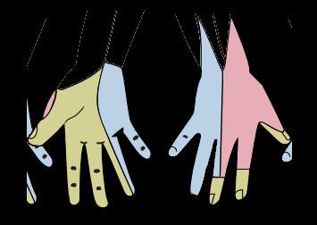
    The median nerve innervates the skin of the palmar side of the index, the thumb and middle finger, half the ring finger, and the nail bed. The lateral part of the palm is supplied by the palmar cutaneous branch of the median nerve, which leaves the nerve proximal to the wrist creases. This palmar cutaneous branch travels in a separate fascial groove adjacent to the flexor carpi radialis and then superficial to the flexor retinaculum. It is therefore spared in carpal tunnel syndrome.

    The muscles of the hand supplied by the median nerve can be remembered using the mnemonic, "LOAF" for Lumbricals 1 & 2, Opponens pollicis, Abductor pollicis brevis and Flexor pollicis brevis. (NB: OAF are the thenar eminence)

    Injury

    Injury of median nerve at different levels causes different syndromes with varying motor and sensory deficits.

    Above the elbow

  • Common mechanism of injury: A supracondylar humerus fracture
  • Motor deficit:
  • Loss of pronation of forearm, weakness in flexion of the hand at the wrist, loss of flexion of radial half of digits and thumb, loss of abduction and opposition of thumb.
  • Presence of an ape hand deformity when the hand is at rest, due to an hyperextension of index finger and thumb, and an adducted thumb.
  • Presence of benediction sign when attempting to form a fist, due to loss of flexion of radial half of digits.
  • Sensory deficit: Loss of sensation in lateral 3 12 digits including their nail beds, and the thenar area.
  • At the elbow

  • Entrapment at the level of the elbow or the proximal forearm could be due to the pronator teres syndrome.
  • Within the proximal forearm: Anterior interosseous syndrome

  • Injury to the anterior interosseous branch in the forearm causes the anterior interosseous syndrome.
  • Common mechanisms: Tight cast, forearm bone fracture
  • Motor deficit: Loss of pronation of forearm, loss of flexion of radial half of digits and thumb.
  • Sensory deficit: None
  • At the wrist

  • Common mechanism: Wrist laceration
  • Motor deficit:
  • Weakness in flexion of radial half of digits and thumb, loss of abduction and opposition of thumb.
  • Presence of an ape hand deformity when the hand is at rest may be likely, due to an hyperextension of index finger and thumb, and an adducted thumb. Nevertheless, an ape hand deformity is not a requirement for a carpal tunnel syndrome diagnosis.
  • Presence of benediction sign when attempting to form a fist, due to weakness in flexion of radial half of digits.
  • Sensory deficit: Loss of sensation in lateral 3 12 digits including their nail beds, and the thenar area.
  • Within the wrist: Carpal tunnel syndrome

  • Common mechanism: Carpal tunnel syndrome, an injury by compression in the carpal tunnel, without transection of the median nerve, due to overuse by activities such as keyboard typing and cooking.
  • Motor deficit:
  • Weakness in flexion of radial half of digits and thumb, weakness in abduction and opposition of thumb.
  • Presence of an ape hand deformity or when attempting to form a fist, the benediction sign, due to compression of the median nerve, as opposed to complete median nerve palsy.
  • Sensory deficit: Numbness and tingling in lateral 3 12 digits including their nail beds but excluding the thenar eminence which is supplied by the palmar cutaneous branch of the median nerve . Unlike in wrist laceration, there is still sensation in the area of the central palm. Sensation is not lost because the palmar cutaneous branch runs above the flexor retinaculum, and is not affected in compression in carpal tunnel syndrome.
  • References

    Median nerve Wikipedia