
Latin nervus ulnaris TA A14.2.03.040 | Dorlands/Elsevier n_05/12566994 | |
 | ||
Innervates flexor carpi ulnarisflexor digitorum profunduslumbrical musclesopponens digiti minimiflexor digiti minimiabductor digiti minimiinterosseiadductor pollicis MeSH A08.800.800.720.050.850 | ||
In human anatomy, the ulnar nerve is a nerve that runs near the ulna bone. The ulnar collateral ligament of elbow joint is in relation with the ulnar nerve. The nerve is the largest unprotected nerve in the human body (meaning unprotected by muscle or bone), so injury is common. This nerve is directly connected to the little finger, and the adjacent half of the ring finger, innervating the palmar side of these fingers, including both front and back of the tips, perhaps as far back as the fingernail beds.
Contents
This nerve can cause an electric shock-like sensation by striking the medial epicondyle of the humerus from posteriorly, or inferiorly with the elbow flexed. The ulnar nerve is trapped between the bone and the overlying skin at this point. This is commonly referred to as bumping one's "funny bone". This name is thought to be a pun, based on the sound resemblance between the name of the bone of the upper arm, the "humerus" and the word "humorous". Alternatively, according to the Oxford English Dictionary it may refer to "the peculiar sensation experienced when it is struck".
Structure
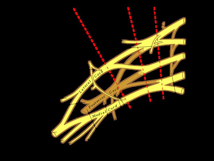
The ulnar nerve originates from the C8-T1 nerve roots (and occasionally carries C7 fibres) which form part of the medial cord of the brachial plexus, and descends on the posteromedial aspect of the humerus. It runs inferior to the posteromedial aspects of the humerus, passing behind the medial epicondyle (in the cubital tunnel) at the elbow where it is exposed for several centimetres.
Forearm
It enters the anterior (flexor) compartment of the forearm between the humeral and ulnar heads of flexor carpi ulnaris, lying under the aponeurosis of flexor carpi ulnaris alongside the ulna. There it supplies one and a half muscles (flexor carpi ulnaris and the medial half of flexor digitorum profundus) and courses with the ulnar artery, travelling inferiorly with it deep to the flexor carpi ulnaris muscle.
In the forearm it gives off the following branches:
Hand
After it travels down the ulna, the ulnar nerve enters the palm of the hand by the Guyon's canal. The ulnar nerve and artery pass superficial to the flexor retinaculum, via the ulnar canal.
Here it gives off the following branches:
The course of the ulnar nerve through the wrist contrasts with that of the median nerve, which travels deep to the flexor retinaculum of the hand.
Sensory
The ulnar nerve also provides sensory innervation to the fifth digit and the medial half of the fourth digit, and the corresponding part of the palm:
Motor
The ulnar nerve and its branches innervate the following muscles in the forearm and hand:
An Articular branch that passes to the elbow joint while the ulnar nerve is passing between the olecranon and medial epicondyle of the humerus.
Clinical significance
The ulnar nerve can suffer injury anywhere between its proximal origin of the brachial plexus all the way to its distal branches in the hand. It is the most commonly injured nerve around the elbow. Although it can be damaged under various circumstances, it is commonly injured by local trauma or physical impingement ("pinched nerve"). Injury of the ulnar nerve at different levels causes specific motor and sensory deficits:
At the elbow
At the wrist
In severe cases, surgery may be performed to relocate or "release" the nerve to prevent further injury.