
Synonyms pulmonary oedema ICD-9-CM 514 518.4 MedlinePlus 000140 | ICD-10 J81 DiseasesDB 11017 | |
 | ||
Specialty Cardiology, critical care medicine | ||
Pulmonary edema is fluid accumulation in the tissue and air spaces of the lungs. It leads to impaired gas exchange and may cause respiratory failure. It is due to either failure of the left ventricle of the heart to remove blood adequately from the pulmonary circulation (cardiogenic pulmonary edema), or an injury to the lung parenchyma or vasculature of the lung (noncardiogenic pulmonary edema). Treatment is focused on three aspects: firstly improving respiratory function, secondly, treating the underlying cause, and thirdly avoiding further damage to the lung. Pulmonary edema, especially acute, can lead to fatal respiratory distress or cardiac arrest due to hypoxia. It is a cardinal feature of congestive heart failure. The term is from the Greek οἴδημα(oídēma, “swelling”), from οἰδέω (oidéō, “I swell”).
Contents
Signs and symptoms
The most common symptom of pulmonary edema is difficulty breathing, but may include other symptoms such as coughing up blood (classically seen as pink, frothy sputum), excessive sweating, anxiety, and pale skin. Shortness of breath can manifest as orthopnea (inability to lie down flat due to breathlessness) and/or paroxysmal nocturnal dyspnea (episodes of severe sudden breathlessness at night). These are common presenting symptoms of chronic pulmonary edema due to left ventricular failure. The development of pulmonary edema may be associated with symptoms and signs of "fluid overload"; this is a non specific term to describe the manifestations of left ventricular failure on the rest of the body and includes peripheral edema (swelling of the legs, in general, of the "pitting" variety, wherein the skin is slow to return to normal when pressed upon), raised jugular venous pressure and hepatomegaly, where the liver is enlarged and may be tender or even pulsatile. Other signs include end-inspiratory crackles (sounds heard at the end of a deep breath) on auscultation and the presence of a third heart sound.
Causes
Pulmonary edema is an accumulation of fluid within the parenchyma and air spaces of the lungs. Classically it is cardiogenic (left ventricular) but fluid may also accumulate due to damage to the lung. This damage may be direct injury or injury mediated by high pressures within the pulmonary circulation. When directly or indirectly caused by increased left ventricular pressure pulmonary edema may form when mean pulmonary pressure rises from the normal of 15 mmHg to above 25 mmHg. Broadly, the causes of pulmonary edema can be divided into cardiogenic and non-cardiogenic. By convention cardiogenic refers to left ventricular causes.
Cardiogenic
Non-cardiogenic
Other
Injury to the lung may also cause pulmonary edema through injury to the vasculature and parenchyma of the lung. The acute lung injury-acute respiratory distress syndrome (ALI-ARDS) covers many of these causes, but they may include:
Some causes of pulmonary edema are less well characterised and arguably represent specific instances of the broader classifications above.
Flash pulmonary edema
Flash pulmonary edema (FPE), is rapid onset pulmonary edema. It is most often precipitated by acute myocardial infarction or mitral regurgitation, but can be caused by aortic regurgitation, heart failure, or almost any cause of elevated left ventricular filling pressures. Treatment of FPE should be directed at the underlying cause, but the mainstays are ensuring adequate oxygenation, diuresis, and decrease of pulmonary circulation pressures.
Recurrence of FPE is thought to be associated with hypertension and may signify renal artery stenosis. Prevention of recurrence is based on managing hypertension, coronary artery disease, renovascular hypertension, and heart failure.
Diagnosis
There is no one single test for confirming that breathlessness is caused by pulmonary edema; indeed, in many cases, the cause of shortness of breath is probably multifactorial.
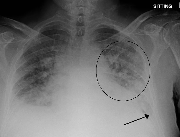
Low oxygen saturation and disturbed arterial blood gas readings support the proposed diagnosis by suggesting a pulmonary shunt. Chest X-ray will show fluid in the alveolar walls, Kerley B lines, increased vascular shadowing in a classical batwing peri-hilum pattern, upper lobe diversion (increased blood flow to the superior parts of the lung), and possibly pleural effusions. In contrast, patchy alveolar infiltrates are more typically associated with noncardiogenic edema
Lung ultrasound, employed by a healthcare provider at the point of care, is also a useful tool to diagnose pulmonary edema; not only is it accurate, but it may quantify the degree of lung water, track changes over time, and differentiate between cardiogenic and non-cardiogenic edema.
Especially in the case of cardiogenic pulmonary edema, urgent echocardiography may strengthen the diagnosis by demonstrating impaired left ventricular function, high central venous pressures and high pulmonary artery pressures.
Blood tests are performed for electrolytes (sodium, potassium) and markers of renal function (creatinine, urea). Liver enzymes, inflammatory markers (usually C-reactive protein) and a complete blood count as well as coagulation studies (PT, aPTT) are also typically requested. B-type natriuretic peptide (BNP) is available in many hospitals, sometimes even as a point-of-care test. Low levels of BNP (<100 pg/ml) suggest a cardiac cause is unlikely.
Prevention
In those with underlying heart disease, effective control of congestive symptoms prevents pulmonary edema.
Dexamethasone is in widespread use for the prevention of high altitude pulmonary edema. Sildenafil is used as a preventive treatment for altitude-induced pulmonary edema and pulmonary hypertension, the mechanism of action is via phosphodiesterase inhibition which raises cGMP, resulting in pulmonary arterial vasodilation and inhibition of smooth muscle cell proliferation. While this effect has only recently been discovered, sildenafil is already becoming an accepted treatment for this condition, in particular in situations where the standard treatment of rapid descent has been delayed for some reason.
Management
The initial management of pulmonary edema, irrespective of the type or cause, is supporting vital functions. Therefore, if the level of consciousness is decreased it may be required to proceed to tracheal intubation and mechanical ventilation to prevent airway compromise. Hypoxia (abnormally low oxygen levels) may require supplementary oxygen, but if this is insufficient then again mechanical ventilation may be required to prevent complications. Treatment of the underlying cause is the next priority; pulmonary edema secondary to infection, for instance, would require the administration of appropriate antibiotics.
Cardiogenic pulmonary edema
Acute cardiogenic pulmonary edema often responds rapidly to medical treatment. Positioning upright may relieve symptoms. Loop diuretics such as furosemide or bumetanide are administered, often together with morphine or diamorphine to reduce respiratory distress. Both diuretics and morphine may have vasodilator effects, but specific vasodilators may be used (particularly intravenous glyceryl trinitrate or ISDN) provided the blood pressure is adequate.
Continuous positive airway pressure and bilevel positive airway pressure (BIPAP/NIPPV) has been demonstrated to reduce the need of mechanical ventilation in people with severe cardiogenic pulmonary edema, and may reduce mortality.
It is possible for cardiogenic pulmonary edema to occur together with cardiogenic shock, in which the cardiac output is insufficient to sustain an adequate blood pressure. This can be treated with inotropic agents or by intra-aortic balloon pump, but this is regarded as temporary treatment while the underlying cause is addressed.