
 | ||
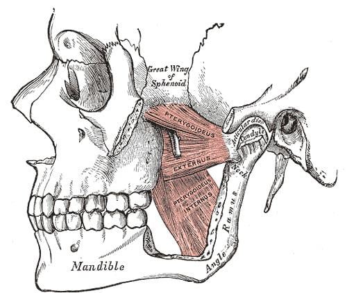
The pterygomandibular space is a fascial space of the head and neck (sometimes also termed fascial spaces or tissue spaces). It is a potential space in the head and is paired on each side. It is located between the medial pterygoid muscle and the medial surface of the ramus of the mandible. The pterygomandibular space is one of the four compartments of the masticator space.
Contents
Anatomic boundaries
The boundaries of each pterygomandibular space are:
Communications
the communications of each pterygomandibular space are:
Contents
In health, the space contains:
Clinical relevance
The pterygomandibular space is the area where local anesthetic solution is deposited during an inferior alveolar nerve block, a common procedure used to anesthetize the distribution of the inferior alveolar nerve. Rarely, pathogenic micro-organisms from the mouth may be seeded into the pterygomandibular space during this injection and cause a needle tract infection of the space. It is also occasionally reported that the needle breaks off and is retained in the pterygomandibular space during this injection. Minor oral surgery is then required to remove the fractured needle. Due to its high vascularity, injections into the pterygomandibular space carry a high risk of intravascular injection (injecting into a blood vessel). Another possible complication of an inferior alveolar nerve block occurs when the needle is placed too deep, passing through the pterygomandibular space and into the parotid gland behind. Branches of the facial nerve (which gives the motor supply to the muscles of facial expression) run through the substance of the parotid gland and so this is manifest as a transient facial palsy. The pterygomandibular space is one of the possible spaces into which a tooth may be displaced into during dental extraction, e.g. of a maxillary wisdom tooth. A mandibular fracture in the angle region may also be the cause of a pterygomandibular space infection.
The signs and symptoms of an isolated pterygomandiublar infection may include trismus (difficulty opening the mouth), however there is not usually any externally visible facial swelling. Intra-orally, there may be swelling and erythema (redness) or the anterior tonsillar pillar (the Palatoglossal arch) and deviation of the uvula to the unaffected side. The airway may be compressed. Treatment is by surgical incision and drainage, and the incision may be placed inside the mouth or two incisions may be used, one inside the mouth and one outside.
Odontogenic infections
Odontogenic infections may spread to involve the pterygomandibular space, and the most common teeth responsible for this are the mandibular second and third molar teeth.