
Entrez 5627 | Ensembl ENSG00000184500 | |
 | ||
Aliases PROS1, PROS, PS21, PS22, PS23, PS24, PS25, PSA, THPH5, THPH6, protein S (alpha) External IDs OMIM: 176880 MGI: 1095733 HomoloGene: 264 GeneCards: PROS1 | ||
Protein S (also known as S-Protein) is a vitamin K-dependent plasma glycoprotein synthesized in the liver. In the circulation, Protein S exists in two forms: a free form and a complex form bound to complement protein C4b-binding protein (C4BP). In humans, protein S is encoded by the PROS1 gene.
Contents
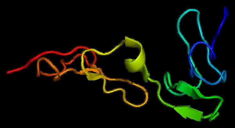
Structure
Protein S is partly homologous to other vitamin K-dependent plasma coagulation proteins, such as protein C and factors VII, IX, and X. Similar to them, it has a Gla domain and several EGF-like domains (four rather than two), but no serine protease domain. Instead, there is a large C-terminus domain that is homologous to plasma steroid hormone-binding proteins such as sex hormone-binding globulin and corticosteroid-binding globulin. It may play a role in the protein functions as either a cofactor for activated protein C (APC) or in binding C4BP.
Additionally, protein S has a peptide between the Gla domain and the EGF-like domain, that is cleaved by thrombin. The Gla and EGF-like domains stay connected after the cleavage by a disulfide bond. However, protein S loses its function as an APC cofactor following either this cleavage or binding C4BP.
Function
The best characterized function of Protein S is its role in the anti coagulation pathway, where it functions as a cofactor to Protein C in the inactivation of Factors Va and VIIIa. Only the free form has cofactor activity.
Protein S can bind to negatively charged phospholipids via the carboxylated Gla domain. This property allows Protein S to function in the removal of cells which are undergoing apoptosis. Apoptosis is a form of cell death that is used by the body to remove unwanted or damaged cells from tissues. Cells, which are apoptotic (i.e. in the process of apoptosis), no longer actively manage the distribution of phospholipids in their outer membrane and hence begin to display negatively charged phospholipids, such as phosphatidyl serine, on the cell surface. In healthy cells, an ATP (Adenosine triphosphate)-dependent enzyme removes these from the outer leaflet of the cell membrane. These negatively charged phospholipids are recognized by phagocytes such as macrophages. Protein S can bind to the negatively charged phospholipids and function as a bridging molecule between the apoptotic cell and the phagocyte. The bridging property of Protein S enhances the phagocytosis of the apoptotic cell, allowing it to be removed 'cleanly' without any symptoms of tissue damage such as inflammation occurring.
Protein S also binds to the nascent complement complex C5,6,7 and prevents this complex from inserting into a membrane. This function prevents the inappropriate activation of the complement system, which would cause uncontrolled systemic inflammation. In fact, Protein S was first discovered in 1977 in this role and it is named after the membrane site that it occupies in the complex.
Pathology
Mutations in the PROS1 gene can lead to Protein S deficiency which is a rare blood disorder which can lead to an increased risk of thrombosis.
Interactions
Protein S has been shown to interact with Factor V.