
Group Group II (ssDNA) | Scientific name Porcine circovirus Rank Species | |
 | ||
Similar Porcine reproductive and respi, Circoviridae, Porcine parvovirus, Mycoplasma hyopneumoniae, Parvovirus | ||
Porcine circovirus
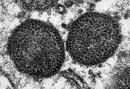
Porcine circovirus (PCV) is a single-stranded DNA virus (class II), that is nonenveloped with an unsegmented circular genome. The viral capsid is icosahedral and approximately 17 nm in diameter. PCV is a member of the virus family Circoviridae.
Contents
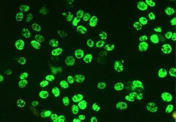
PCVs are the smallest viruses replicating autonomously in eukaryotic cells. They replicate in the nucleus of infected cells, using the host polymerase for genome amplification.
There are 2 strains: type 1 and type 2. Porcine Circovirus Associated Disease is caused by porcine circovirus type 2 (PCV2).
PCV-1 (first identified in 1974) readily infects, but is not known to cause disease in swine; the type 2 has caused problems in recent years with the increasing occurrence of postweaning multisystemic wasting syndrome (PMWS), which over time results in significant depletion of lymphocytes; postmortem examination of diseased animals reveals enlarged lymph nodes and abnormal lung tissue.
PCV-2 (first isolated in 1997) causes PMWS. However, viral infection by itself tends to cause only mild disease, and co-factors such as other infections or immunostimulation seem necessary for development of severe disease. For example, concurrent infection with porcine parvovirus or PRRS virus, or immunostimulation lead to increased replication of PCV-2 and more severe disease in PCV-2-infected pigs.
PCV-1 and PCV-2 show a high degree of sequence identity and a similar genomic organisation; nevertheless, the basis of the distinct pathogenicity has not yet been unravelled.
An effective vaccination is now available. Fort Dodge Animal Health (Wyeth) launched the first USDA approved vaccine in 2006, containing an inactivated virus (ATCvet code: QI09AA07 (WHO)).
Genome
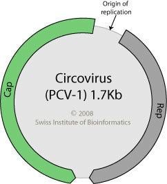
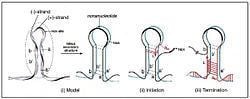
PCV's genome is one of the simplest of all viruses, requiring only a capsid protein (ORF2) and two replicase proteins (ORF1) in order to replicate and produce a functional virus. Due to the simplicity of PCV, it must rely heavily on the host's cellular machinery to replicate. The origin of replication is located on a small octanucleotide stem-loop that is flanked by palindromic repeats, with the ORF's being located head-to-head on both sides of the Ori. Specifically, ORF1 is located clockwise and ORF2 is located counterclockwise of the Ori. The two replicase enzymes that are created from ORF1, Rep and Rep', are conserved between the two types of PCV, and are part of the early phase of the virus. The replicases differ in that Rep is the full ORF1 transcript of 312 amino acids, whereas Rep' is a truncated form of ORF1 as a result of splicing and is only 168 amino acids in length. The promoter for rep (Prep) contains an Interferon-Stimulated Response Element (ISRE) that suggests Rep and Rep' are regulated by cytokine involvement, and is probably a means for the virus to overcome the host's immune responses to infection. Rep and Rep' form a dimer that binds to two hexameric regions adjacent to the stem-loop, H1 and H2, which is required for replication. When the dimer binds to this region, the replicases cleave the loop region of the stem-loop and remain covalently bound to the H1 and H2 regions of the DNA, which becomes the 5' end of the DNA. The newly formed 3'OH end forms a primer using host RNA polymerase, which is then used by the host's DNA polymerase to begin transcription of the viral DNA via rolling circle replication. After the complementary DNA strand has been created, the stem region of the stem-loop forms a loose, non-hydrogen bonded, quadruplet DNA structure. This loosely associated structure can form short lived DNA-trimers which forms two templates for replication, as well as maintaining the nucleic integrity of the stem region of the stem-loop. The termination of the replication sequence has not been identified, yet, though there is evidence supporting that Rep also represses its own promoter, Prep.
The ORF2 region encodes Cap, which differs slightly between PCV-1 and PCV-2. This variation within PCV may explain why PCV-1 is non-pathogenic, while PCV-2 is pathogenic. The promoter for this protein is located within ORF1, within the site where Rep' is truncated, and is splice from the same exon to the starting point of the ORF2 coding region and expressed during both early and late phases. This is the immunogenic region of the virus and is the primary area of research for creating a vaccine to treat PMWS.
There is a third gene encoded in the opposite orientation to ORF1 in the genome. This gene is transcribed and is an essential gene involved in viral replication.
DNA
It is a replicating entity with one of the smallest DNA strands consisting of a simple loop of DNA. Its DNA sequence is 1768 base pairs long:
1 accagcgcac ttcggcagcg gcagcacctc ggcagcacct cagcagcaac atgcccagca 61 agaagaatgg aagaagcgga ccccaaccac ataaaaggtg ggtgttcacg ctgaataatc 121 cttccgaaga cgagcgcaag aaaatacggg agctcccaat ctccctattt gattatttta 181 ttgttggcga ggagggtaat gaggaaggac gaacacctca cctccagggg ttcgctaatt 241 ttgtgaagaa gcaaactttt aataaagtga agtggtattt gggtgcccgc tgccacatcg 301 agaaagccaa aggaactgat cagcagaata aagaatattg cagtaaagaa ggcaacttac 361 ttattgaatg tggagctcct cgatctcaag gacaacggag tgacctgtct actgctgtga 421 gtaccttgtt ggagagcggg agtctggtga ccgttgcaga gcagcaccct gtaacgtttg 481 tcagaaattt ccgcgggctg gctgaacttt tgaaagtgag cgggaaaatg cagaagcgtg 541 attggaagac caatgtacac gtcattgtgg ggccacctgg gtgtggtaaa agcaaatggg 601 ctgctaattt tgcagacccg gaaaccacat actggaaacc acctagaaac aagtggtggg 661 atggttacca tggtgaagaa gtggttgtta ttgatgactt ttatggctgg ctgccgtggg 721 atgatctact gagactgtgt gatcgatatc cattgactgt agagactaaa ggtggaactg 781 tacctttttt ggcccgcagt attctgatta ccagcaatca gaccccgttg gaatggtact 841 cctcaactgc tgtcccagct gtagaagctc tctatcggag gattacttcc ttggtatttt 901 ggaagaatgc tacagaacaa tccacggagg aagggggcca gttcgtcacc ctttcccccc 961 catgccctga atttccatat gaaataaatt actgagtctt ttttatcact tcgtaatggt 1021 ttttattttt catttagggg ttaagtgggg ggtctttaag attaaattct ctgaattgta 1081 catacatggt tacacggata ttgtagtcct ggtcgtattt actgttttcg aacgcagtgc 1141 cgaggcctac gtggtccaca tttctagagg tttgtagcct cagccaaagc tgattccttt 1201 tgttatttgg ttggaagtaa tcaatagtgg agtcaagaac aggtttgggt gtgaagtaac 1261 gggagtggta ggagaagggt tgggggattg tatggcggga ggagtagttt acatatgggt 1321 cataggttag ggcattggcc tttggtacaa agttatcatc tagaataaca gcagtggagc 1381 ccactcccct atcaccctgg gtgatggggg agcagggcca gaattcaacc ttaacttttc 1441 ttattctgta gtattcaaag ggtatagaga ttttgttggt cccccctccc gggggaacaa 1501 agtcgtcaag cttaaatctc atcatgtcca ccgcccagga gggcgttgtg actgtggtac 1561 gcttgacagt atatccgaag gtgcgggaga ggcgggtgtt gaagatgcca tttttccttc 1621 tccaacggta gcggtggcgg gggtggacga gccaggggcg gcggcggagg atctggccaa 1681 gatggctgcg ggggcggtgt cttcttctgc ggtaacgcct ccttggatac gtcatagctg 1741 aaaacgaaag aagtgcgctg taagtattEntry
PCV infects a wide variety of cell types, including hepatocytes, cardiomyocytes, and macrophages. However, until recently, it was unknown exactly how attachment and entry into these cells was achieved. Research has shown that PCV utilizes clathrin-mediated endocytosis to enter the cell, though it's stipulated that there may still be other factors that haven't been identified. Once endocytosed, the endosome and lysosome formation causes an acidic pH shift, which allows ATP-driven uncoating of the virus and allows it to escape the endosomes and lysosomes. After the virus escapes the endosomes and lysosomes, it travels to the nucleus through unknown means.
Escape
Besides ORF1 and ORF2, there is also an ORF3 which is not necessarily required for PCV to survive within the host. Research has shown that the protein coded in ORF3 can modulate the host cell's cell-division cycle and cause cell-mediated, virus-induced apoptosis. Using a yeast two-hybrid screening system of ORF3 against the porcine cDNA library indicated that the ORF3 protein interacts with the porcine pPirh2, which is an E3 ubiquitin ligase. This E3 ubiquitin ligase normally interacts with p53 during the cell division cycle and prevents it from halting the cell division cycle at S-phase. However, ORF3 also interacts with pPirh2 at the same region as p53 and causes an upregulation of p53 expression. This increase in p53 stops the cell division cycle and the result of this is p53 mediated apoptosis, which releases PCV into the extracellular environment.
Porcine circovirus DNA identified in human vaccine
On 22 March 2010, U.S. Food and Drug Administration (FDA) health authorities recommended doctors suspend using Rotarix, one of two vaccines licensed in the United States against rotavirus, due to findings of viral DNA contamination. An independent academic research team in San Francisco identified porcine circovirus-1 DNA in two lots of Rotarix, and follow-up work by GlaxoSmithKline confirmed the contamination in working cells and the viral "seed" used Rotarix production, also confirming the material was likely present since the early stages of product development, including the clinical trials for FDA approval.
Testing of the other licensed vaccine against rotavirus infection, RotaTeq, has also detected some components of both PCV-1 and PCV-2. Porcine circovirus 1 is not known to cause disease in humans or other animals.
As of 8 June 2010, FDA has, based on a careful review of a variety of scientific information, determined it is appropriate for clinicians and public health professionals in the United States to use both Rotarix and RotaTeq vaccine.