
Code TH H3.04.03.0.00020 TH H3.04.03.0.00020 | TA A05.6.01.014 FMA 76466 | |
 | ||
Latin noduli lymphoidei aggregati | ||
Peyer's patches (or aggregated lymphoid nodules, or occasionally PP for brevity) are organized lymphoid follicles, named after the 17th-century Swiss anatomist Johann Conrad Peyer. They are an important part of gut associated lymphoid tissue usually found in humans in the lowest portion of the small intestine, mainly in the distal jejunum and the ileum, but also could be detected in the duodenum.
Contents
Structure
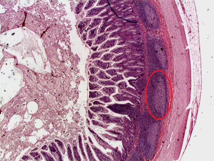
Peyer's patches are observable as elongated thickenings of the intestinal epithelium measuring a few centimeters in length. About 100 are found in humans. Microscopically, Peyer's patches appear as oval or round lymphoid follicles (similar to lymph nodes) located in the submucosa layer of the ileum and extend into the mucosa layer. The number of Peyer's patches peaks at age 15–25 and then declines during adulthood. In the distal ileum, they are numerous and they form a lymphoid ring. At least 46% of Peyer's patches are concentrated in the distal 25 cm of ileum in humans. It is important to note that there are large variations in size, shape, and distribution of Peyer's patches from one individual to another one. In adults, B lymphocytes are seen to dominate the follicles' germinal centers. T lymphocytes are found in the zones between follicles. Among the mononuclear cells, CD4+/CD25+ (10%) cells and CD8+/CD25+ (5%) cells are more abundant in Peyer's patches than in the peripheral blood.
Peyer's patches are characterized by the follicle-associated epithelium (FAE), which covers every lymphoid follicles. FAE differs from typical small intestinal villus epithelium: it has fewer goblet cells therefore mucus layer is thinner, and it is also characterized by the presence of specialized M cells or microfold cells, which provide uptake and transport of antigens from lumen. Moreover, basal lamina of follicle-associated epithelium is more porous compare to intestinal villus. Finally, follicle-associated epithelium is less permeable for ions and macromolecules, basically due to higher expression of tight junction proteins.
Function
Because the lumen of the gastrointestinal tract is exposed to the external environment, much of it is populated with potentially pathogenic microorganisms. Peyer's patches thus establish their importance in the immune surveillance of the intestinal lumen and in facilitating the generation of the immune response within the mucosa.
Pathogenic microorganisms and other antigens entering the intestinal tract encounter macrophages, dendritic cells, B-lymphocytes, and T-lymphocytes found in Peyer's patches and other sites of gut-associated lymphoid tissue (GALT). Peyer's patches thus act for the gastrointestinal system much as the tonsils act for the respiratory system, trapping foreign particles, surveilling them, and destroying them.
Peyer's patches are covered by a special follicle-associated epithelium that contains specialized cells called microfold cells (M cells) which sample antigen directly from the lumen and deliver it to antigen-presenting cells (located in a unique pocket-like structure on their basolateral side). Dendritic cells and macrophages can also directly sample the lumen by extending dendrites through transcellular M cell-specific pores. At the same time the paracellular pathway of follicle-associated epithelium is closed tightly to prevent penetration of antigens and continuous contact with immune cells. T cells, B-cells and memory cells are stimulated upon encountering antigen in Peyer's patches. These cells then pass to the mesenteric lymph nodes where the immune response is amplified. Activated lymphocytes pass into the blood stream via the thoracic duct and travel to the gut where they carry out their final effector functions.The maturation of B-lymphocytes takes place in the Peyer's patch.
Clinical significance
Although important in the immune response, excessive growth of lymphoid tissue in Peyer's patches is pathologic, as hypertrophy of Peyer's patches has been closely associated with idiopathic intussusception.
The hypertrophy of Peyer's patches has also been associated with susceptibility to transmissible spongiform encephalopathies (commonly known as prion diseases).
Salmonella typhi and poliovirus also target this section of the intestine.