
 | ||
An organoid is a miniaturized and simplified version of an organ produced in vitro in three dimensions that shows realistic micro-anatomy. They are derived from one or a few cells from a tissue, embryonic stem cells or induced pluripotent stem cells, which can self-organize in three-dimensional culture owing to their self-renewal and differentiation capacities. The technique for growing organoids has rapidly improved since the early 2010s, and it was named by The Scientist as one of the biggest scientific advancements of 2013.
Contents
History
Attempts to create organs ‘’in vitro’’ started with one of the first dissociation- reaggregation experiment where Henry Van Peters Wilson demonstrated that mechanically dissociated sponge cells can reaggregate and self-organize to generate a whole organism. In the subsequent decades, multiple labs were able to generate different types of organs ‘’in vitro’’ through the dissociation and reaggregation of organ tissues obtained from amphibians and embryonic chicks. The phenomena of mechanically dissosciated cells aggregating and reorganizing to reform the tissue they were obtained from subsequently led to the development of the Differential adhesion hypothesis by Malcolm Steinberg. With the advent of the field of stem cell biology, the potential of stem cells in to form organs in ‘’vitro’’ was realized early on with the observation that when stem cells form teratomas or embryoid body, the differentiated cells can organize into different structures resembling those found in multiple tissue types. The advent of the field of organoids, started with a shift from culturing and differentiating stem cells in 2D media, to 3D media to allow for the development of the complex 3-dimensional structures of organs. Since 1987, researchers have devised different methods for 3-D culturing, and were able to utilize different types of stem cells to generate organoids resembling a multitude of organs. In 2008, Yoshiki Sasai and his team at RIKEN institute demonstrated that stem cells can be coaxed into balls of neural cells that self-organize into distinctive layers. In 2009 the Laboratory of Hans Clevers at Hubrecht Institute and University Medical Center Utrecht, The Netherlands showed that single LGR5 stem cells build crypt-villus structures in vitro without a mesenchymal niche. In 2010, Mathieu Unbekandt & Jamie A. Davies demonstrated the production of renal organoids from murine fetus-derived renogenic stem cells: subsequent reports showed significant physiological function of these organoids in vitro and in vivo.
In 2013, Madeline Lancaster at the Austrian Academy of Sciences established a protocol for culturing cerebral organoids derived from stem cells that mimic the developing human brain's cellular organization. In 2014, Artem Shkumatov et al. at the University of Illinois at Urbana-Champaign demonstrated that cardiovascular organoids can be formed from ES cells through modulation of the substrate stiffness, to which they adhere. Physiological stiffness promoted three-dimensionality of EBs and cardiomyogenic differentiation.
Takebe et al. demonstrate a generalized method for organ bud formation from diverse tissues by combining pluripotent stem cell-derived tissue-specific progenitors or relevant tissue samples with endothelial cells and mesenchymal stem cells. They suggested that the less mature tissues, or organ buds, generated through the self-organized condensation principle might be the most efficient approach toward the reconstitution of mature organ functions after transplantation, rather than condensates generated from cells of a more advanced stage
Properties
Lancaster and Knoblich define an organoid as a collection of organ-specific cell types that develops from stem cells or organ progenitors, self-organizes through cell sorting and spatially restricted lineage commitment in a manner similar to in vivo, and exhibits the following properties:
Process
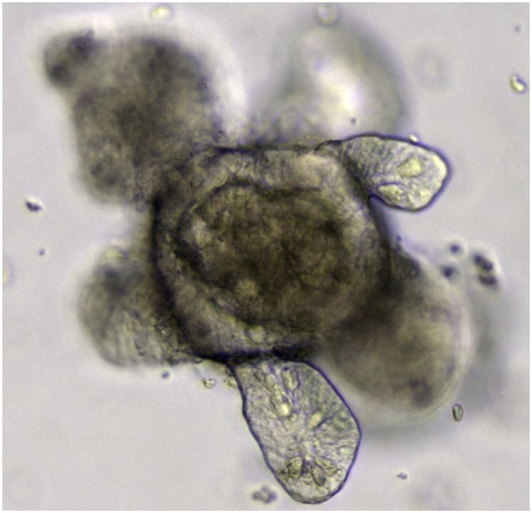
Organoid formation generally requires culturing the stem cells or progenitor cells in a 3D media. The 3D media can be made using an extracellular matrix hydrogel Matrigel, which is a laminin- rich extracellular matrix that is secreted by the Engelbreth- Holm-Swarm tumor line. Organoid bodies can then be made through embedding stem cells in the 3D media. When pluripotent stem cells are used for the creation of the organoid, the cells are usually, but not all the time, allowed to form embryoid bodies. Those embryoid bodies are then pharmacologically treated with patterning factors to drive the formation of the desired organoid identity. Organoids have also been created using adult stem cells extracted from the target organ, and cultured in 3D media
Types of organoids
A multitude of organ structures have been recapitulated using organoids. This section aims to outline the state of the field as of now through providing an abridged list of the organoids that have been successfully created, along with a brief outline based on the most recent literature for each organoid, and examples of how it has been utilized in research.
Basic research with organoids
Organoids are an excellent tool to study basic biological processes. Organoids enable to study how cells interact together in an organ, their interaction with their environment, how diseases affect them and the effect of drugs. In vitro culture makes this system easy to manipulate and facilitates their monitoring. While organs are difficult to culture because their size limits the penetration of nutrients, the small size of organoids limits this problem. On the other hand, they don´t exhibit all organ features and interactions with other organs are not recapitulated in vitro. While research on stem cells and regulation of stemness was the first field of application of intestinal organoids, they are now also used to study e.g. uptake of nutrients, drug transport and secretion of incretin hormones. This is of great relevance in the context of malabsorption diseases as well as metabolic diseases such as obesity, insulin resistance, and diabetes.
Organoid models of disease
Organoids provide an opportunity to create cellular models of human disease, which can be studied in the laboratory to better understand the causes of disease and identify possible treatments. In one example, the genome editing system called CRISPR was applied to human pluripotent stem cells to introduce targeted mutations in genes relevant to two different kidney diseases, polycystic kidney disease and focal segmental glomerulosclerosis. These CRISPR-modified pluripotent stem cells were subsequently grown into human kidney organoids, which exhibited disease-specific phenotypes. Kidney organoids from stem cells with polycystic kidney disease mutations formed large, translucent cyst structures from kidney tubules. Kidney organoids with mutations in a gene linked to focal segmental glomerulosclerosis developed junctional defects between podocytes, the filtering cells affected in that disease. Importantly, these disease phenotypes were absent in control organoids of identical genetic background, but lacking the CRISPR mutations. These experiments demonstrate how organoids can be utilized to create complex models of human disease in the laboratory, which recapitulate tissue-level phenotypes in a petri dish.
Personalised medicine with organoids
Intestinal organoids grown from rectal biopsies using culture protocols established by the Clevers group have been used to model cystic fibrosis, and led to the first application of organoids for personalised treatment. Cystic fibrosis is an inherited disease that is caused by gene mutations of the cystic fibrosis transmembrane conductance regulator gene that encodes an epithelial ion channel necessary for healthy epithelial surface fluids. Studies by the laboratory of Jeffrey Beekman (Wilhelmina Children's Hospital, University Medical Center Utrecht, The Netherlands) described in 2013 that stimulation of colorectal organoids with cAMP-raising agonists such as forskolin or cholera toxin induced rapid swelling of organoids in a fully CFTR dependent manner. Whereas organoids from non-cystic fibrosis subjects swell in response to forskolin as a consequence of fluid transport into the organoids' lumens, this is severely reduced or absent in organoids derived from people with cystic fibrosis. Swelling could be restored by therapeutics that repair the CFTR protein (CFTR modulators), indicating that individual responses to CFTR modulating therapy could be quantitated in a preclinical laboratory setting. Schwank et al. also demonstrated that the intestinal cystic fibrosis organoid phenotype could be repaired by CRISPR-Cas9 gene editing in 2013.
Follow-up studies by Dekkers et al. in 2016 revealed that quantitative differences in forskolin-induced swelling between intestinal organoids derived from people with cystic fibrosis associate with known diagnostic and prognostic markers such as CFTR gene mutations or in vivo biomarkers of CFTR function. In addition, the authors demonstrated that CFTR modulator responses in intestinal organoids with specific CFTR mutations correlated with published clinical trial data of these treatments. This led to preclinical studies where organoids from patients with extremely rare CFTR mutations for who no treatment was registered were found to respond strongly to a clinically available CFTR modulator. The suggested clinical benefit of treatment for these subjects based on the preclinical organoid test was subsequently confirmed upon clinical introduction of treatment by members of the clinical CF center under supervision of Kors van der Ent (Department of Paediatric Pulmonology, Wilhelmina Children's Hospital, University Medical Center Utrecht, The Netherlands). These studies show for the first time that organoids can be used for the individual tailoring of therapy or personalised medicine.
Organoids as a model for developmental biology
Organoids offer researchers an exceptional model to study developmental biology. Since the identification of pluripotent stem cells, there have been great advancements in directing pluripotent stem cells fate ‘’in vitro’’ using 2D cultures. These advancements in PSC fate direction, coupled with the advancements in 3D culturing techniques allowed for the creation of organoids that recapitulate the properties of various specific subregions of a multitude of human organs. The use of these organoids has thus greatly contributed to expanding our understanding of the processes of organogenesis, and the field of developmental biology. In central nervous system development, for example, organoids have contributed to our understanding of the physical forces that underlie retinal cup formation, the species-specific dimensionality of the eye, and the factors underlying the control of progenitor cells in the cortex.