
 | ||
Optogenetics (from Greek optikós, meaning 'seen, visible') is a biological technique which involves the use of light to control cells in living tissue, typically neurons, that have been genetically modified to express light-sensitive ion channels. It is a neuromodulation method employed in neuroscience that uses a combination of techniques from optics and genetics to control and monitor the activities of individual neurons in living tissue—even within freely-moving animals—and to precisely measure these manipulation effects in real-time. The key reagents used in optogenetics are light-sensitive proteins. Neuronal control is achieved using optogenetic actuators like channelrhodopsin, halorhodopsin, and archaerhodopsin, while optical recording of neuronal activities can be made with the help of optogenetic sensors for calcium (GCaMP), vesicular release (synapto-pHluorin), Neurotransmitter (GluSnFRs), or membrane voltage (Arc Lightning, ASAP1). Control or recording is confined to genetically defined neurons and performed in a spatiotemporal-specific manner by light.
Contents
- History
- Description
- Technique
- Issues
- Applications
- Amygdala
- Olfactory Bulb
- Nucleus accumbens
- Prefrontal cortex
- Heart
- Spiral ganglion
- Brainstem
- Precise temporal control of interventions
- Hippocampus
- Additional reading
- References
The earliest approaches for optogenetic control were invented and applied by Boris Zemelman and Gero Miesenböck, at the Sloan-Kettering Cancer Center in New York City, and Dirk Trauner, Richard Kramer and Ehud Isacoff at the University of California, Berkeley; these methods conferred light sensitivity but were never reported to be useful by other laboratories due to the multiple components these approaches required. A distinct single-component approach involving microbial opsin genes introduced in 2005 turned out to be widely applied, as described below. Optogenetics is known for the high spatial and temporal resolution that it provides in altering the activity of specific types of neurons to control a subject's behaviour.
In 2010, optogenetics was chosen as the "Method of the Year" across all fields of science and engineering by the interdisciplinary research journal Nature Methods. At the same time, optogenetics was highlighted in the article on "Breakthroughs of the Decade" in the academic research journal Science. These journals also referenced recent public-access general-interest video Method of the year video and textual SciAm summaries of optogenetics.
History
The "far-fetched" possibility of using light for selectively controlling precise neural activity (action potential) patterns within subtypes of cells in the brain was thought of by Francis Crick in his Kuffler Lectures at the University of California in San Diego in 1999. An earlier use of light to activate neurons was carried out by Richard Fork, who demonstrated laser activation of neurons within intact tissue, although not in a genetically-targeted manner. The earliest genetically targeted method, which used light to control genetically-sensitised neurons, was reported in January 2002 by Boris Zemelman (now at UT Austin) and Gero Miesenböck, who employed Drosophila rhodopsin photoreceptors for controlling neural activity in cultured mammalian neurons. In 2003 Zemelman and Miesenböck developed a second method for light-dependent activation of neurons in which single ionotropic channels TRPV1, TRPM8 and P2X2 were gated by caged ligands in response to light. Beginning in 2004, the Kramer and Isacoff groups developed organic photoswitches or "reversibly caged" compounds in collaboration with the Trauner group that could interact with genetically introduced ion channels. TRPV1 methodology, albeit without the illumination trigger, was subsequently used by several laboratories to alter feeding, locomotion and behavioral resilience in laboratory animals. However, these initial light-based approaches for altering neuronal activity were not applied outside the original laboratories, likely because the easier to employ channelrhodopsin was cloned soon thereafter.
Dr. Zhuo-Hua Pan of Wayne State University, researching on restore sight to blindness, thought about using channelrhodopsin when it came out in late 2003. By February 2004, he was trying channelrhodopsin out in ganglion cells — the neurons in our eyes that connect directly to the brain — that he had cultured in a dish. They became electrically active in response to light. Over the moon with excitement, Pan applied for a grant from the National Institutes of Health. The NIH awarded him $300,000, with the comment that his research was "quite an unprecedented, highly innovative proposal, bordering on the unknown."
In April 2005, Susana Lima and Miesenböck reported the first use of genetically-targeted P2X2 photostimulation to control the behaviour of an animal. They showed that photostimulation of genetically circumscribed groups of neurons, such as those of the dopaminergic system, elicited characteristic behavioural changes in fruit flies. In August 2005, Karl Deisseroth's laboratory in the Bioengineering Department at Stanford including graduate students Ed Boyden and Feng Zhang (both now at MIT) published the first demonstration of a single-component optogenetic system, beginning in cultured mammalian neurons. using channelrhodopsin, a single-component light-activated cation channel from unicellular algae, whose molecular identity and principal properties rendering it useful for optogenetic studies had been first reported in November 2003 by Georg Nagel. The groups of Gottschalk and Nagel were the first to extend the usability of Channelrhodopsin-2 for controlling neuronal activity to the intact animal by showing that motor patterns in the roundworm Caenorhabditis elegans could be evoked by targeted expression and stimulation of Channelrhodopsin-2 in selected neural circuits (published in December 2005). Now optogenetics has been routinely combined with brain region-and cell type-specific Cre/loxP genetic methods developed for Neuroscience by Joe Z. Tsien back in 1990s to activate or inhibit specific brain regions and cell-types in vivo.
The primary tools for optogenetic recordings have been genetically encoded calcium indicators (GECIs). The first GECI to be used to image activity in an animal was cameleon, designed by Atsushi Miyawaki, Roger Tsien and coworkers. Cameleon was first used successfully in an animal by Rex Kerr, William Schafer and coworkers to record from neurons and muscle cells of the nematode C. elegans. Cameleon was subsequently used to record neural activity in flies and zebrafish. In mammals, the first GECI to be used in vivo was GCaMP, first developed by Nakai and coworkers. GCaMP has undergone numerous improvements, and GCaMP6 in particular has become widely used throughout neuroscience.
In 2010 Karl Deisseroth at Stanford University was awarded the inaugural HFSP Nakasone Award "for his pioneering work on the development of optogenetic methods for studying the function of neuronal networks underlying behavior". In 2012 Gero Miesenböck was awarded the InBev-Baillet Latour International Health Prize for "pioneering optogenetic approaches to manipulate neuronal activity and to control animal behaviour." In 2013 Ernst Bamberg, Ed Boyden, Karl Deisseroth, Peter Hegemann, Gero Miesenböck and Georg Nagel were awarded The Brain Prize for "their invention and refinement of optogenetics."
Description
Optogenetics provides millisecond-scale temporal precision which allows the experimenter to keep pace with fast biological information processing (for example, in probing the causal role of specific action potential patterns in defined neurons). Indeed, to probe the neural code, optogenetics by definition must operate on the millisecond timescale to allow addition or deletion of precise activity patterns within specific cells in the brains of intact animals, including mammals (see Figure 1). By comparison, the temporal precision of traditional genetic manipulations (employed to probe the causal role of specific genes within cells, via "loss-of-function" or "gain of function" changes in these genes) is rather slow, from hours or days to months. It is important to also have fast readouts in optogenetics that can keep pace with the optical control. This can be done with electrical recordings ("optrodes") or with reporter proteins that are biosensors, where scientists have fused fluorescent proteins to detector proteins. An example of this is voltage-sensitive fluorescent protein (VSFP2). Additionally, beyond its scientific impact optogenetics represents an important case study in the value of both ecological conservation (as many of the key tools of optogenetics arise from microbial organisms occupying specialized environmental niches), and in the importance of pure basic science as these opsins were studied over decades for their own sake by biophysicists and microbiologists, without involving consideration of their potential value in delivering insights into neuroscience and neuropsychiatric disease.
Light-Activated Membrane Channels
The hallmark of optogenetics therefore is introduction of fast light-activated channels and enzymes that allow temporally precise manipulation of electrical and biochemical events while maintaining cell-type resolution through the use of specific targeting mechanisms. Among the microbial opsins which can be used to investigate the function of neural systems are the channelrhodopsins (ChR2, ChR1, VChR1, and SFOs) to excite neurons and anion-conducting channelrhodopsins for light-induced inhibition. For silencing, halorhodopsin (NpHR), enhanced halorhodopsins (eNpHR2.0 and eNpHR3.0), archaerhodopsin (Arch), Leptosphaeria maculans fungal opsins (Mac), and enhanced bacteriorhodopsin (eBR) have been employed to inhibit neurons (see Figure 2), including in freely-moving mammals.
Moreover, optogenetic control of well-defined biochemical events within behaving mammals is now also possible. Building on prior work fusing vertebrate opsins to specific G-protein coupled receptors a family of chimeric single-component optogenetic tools was created that allowed researchers to manipulate within behaving mammals the concentration of defined intracellular messengers such as cAMP and IP3 in targeted cells Other biochemical approaches to optogenetics (crucially, with tools that displayed low activity in the dark) followed soon thereafter, when optical control over small GTPases and adenylyl cyclases was achieved in cultured cells using novel strategies from several different laboratories. This emerging repertoire of optogenetic probes now allows cell-type-specific and temporally precise control of multiple axes of cellular function within intact animals.
Hardware for Light Application
Another necessary factor is hardware (e.g. integrated fiberoptic and solid-state light sources) to allow specific cell types, even deep within the brain, to be controlled in freely behaving animals. Most commonly, the latter is now achieved using the fiberoptic-coupled diode technology introduced in 2007, though to avoid use of implanted electrodes, researchers have engineered ways to inscribe a "window" made of zirconia that has been modified to be transparent and implanted in mice skulls, to allow optical waves to penetrate more deeply to stimulate or inhibit individual neurons. To stimulate superficial brain areas such as the cerebral cortex, optical fibers or LEDs can be directly mounted to the skull of the animal. More deeply implanted optical fibers have been used to deliver light to deeper brain areas. Complementary to fiber-tethered approaches, completely wireless techniques have been developed utilizing wirelessly delivered power to headborne LEDs for unhindered study of complex behaviors in freely behaving organisms.
Expression of Optogenetic Actuators
Optogenetics also necessarily includes the development of genetic targeting strategies such as cell-specific promoters or other customized conditionally-active viruses, to deliver the light-sensitive probes to specific populations of neurons in the brain of living animals (e.g. worms, fruit flies, mice, rats, and monkeys). In invertebrates such as worms and fruit flies some amount of retinal isomerase all-trans-retinal (ATR) is supplemented with food. A key advantage of microbial opsins as noted above is that they are fully functional without the addition of exogenous co-factors in vertebrates.
Technique
The technique of using optogenetics is flexible and adaptable to the experimenter's needs. For starters, experimenters genetically engineer a microbial opsin based on the gating properties (rate of excitability, refractory period,etc..) required for the experiment.
There is a challenge in introducing the microbial opsin, an optogenetic actuator, into a specific region of the organism in question. A rudimentary approach is to introduce an engineered viral vector that contains the optogenetic actuator gene attached to a recognizable promoter such as CAMKIIα. This allows for some level of specificity as cells that already contain and can translate the given promoter will be infected with the viral vector and hopefully express the optogenetic actuator gene.
Another approach is the creation of transgenic mice where the optogenetic actuator gene is introduced into mice zygotes with a given promoter, most commonly Thy1. Introduction of the optogenetic actuator at an early stage allows for a larger genetic code to be incorporated and as a result, increases the specificity of cells to be infected.
A third and rather novel approach that has been developed is creating transgenic mice with Cre-Recombinase, an enzyme that catalyzes recombination between two lox-P sites. Then by introducing an engineered viral vector containing the optogenetic actuator gene in between two lox-P sites, only the cells containing the Cre-Recombinase will express the microbial opsin. This last technique has allowed for multiple modified optogenetic actuators to be used without the need to create a whole line of transgenic animals every time a new microbial opsin is needed.
After the introduction and expression of the microbial opsin, depending on the type of analysis being performed, application of light can be placed at the terminal ends or the main region where the infected cells are situated. Light stimulation can be performed with a vast array of instruments from light emitting diodes (LEDs) or diode-pumped solid state (DPSS). These light sources are most commonly connected to a computer through a fiber optic cable. Recent advances include the advent of wireless head-mounted devices that also apply LED to targeted areas and as a result give the animal more freedom of mobility to reproduce in vivo results.
Issues
One of the main problems of optogenetics is that not all of the cells in question may express the microbial opsin gene and this is due to each cell's transcriptional and translational mechanisms, and mutations that may occur. Thus, light activation of a population of cells might not be representable of in vivo occurrences. Another problem with the technique is that light stimulation produces a synchronous activation of infected cells and this removes any individual cell properties of activation among the population affected. Therefore, it is difficult to understand how the cells in the population affected communicate with one another or how their phasic properties of activation may relate to the circuitry being observed. An issue with Channelrhodopsin-2 is that its gating properties don't mimic in vivo cation channels of cortical neurons. A solution to this issue with a protein's kinetic property is introduction of variants of Channelrhodopsin-2 with more favorable kinetics.
Applications
The field of optogenetics has furthered the fundamental scientific understanding of how specific cell types contribute to the function of biological tissues such as neural circuits in vivo (see references from the scientific literature below). Moreover, on the clinical side, optogenetics-driven research has led to insights into Parkinson's disease and other neurological and psychiatric disorders. Indeed, optogenetics papers in 2009 have also provided insight into neural codes relevant to autism, Schizophrenia, drug abuse, anxiety, and depression.
Amygdala
Optogenetic approaches have been used to map neural circuits in the amygdala that contribute to fear conditioning. One such example of a neural circuit is the connection made from the basolateral amydala to the dorsal-medial prefrontal cortex where neuronal oscillations of 4 Hz have been observed in correlation to fear induced freezing behaviors in mice. Transgenic mice were introduced with Channelrhodoposin-2 attached with a Parvalbumin-Cre promoter that selectively infected interneurons located both in the basolateral amygdala and the dorsal-medial prefrontal cortex responsible for the 4 Hz oscillations. The interneurons were optically stimulated generating a freezing behavior and as a result provided evidence that these 4 Hz oscillations may be responsible for the basic fear response produced by the neuronal populations along the dorsal-medial prefrontal cortex and basolateral amygdala.
Olfactory Bulb
Optogenetic activation of olfactory sensory neurons was critical for demonstrating timing in odor processing and for mechanism of neuromodulatory mediated olfactory guided behaviors (e.g. aggression, mating) In addition,with the aid of optogenetics, evidence has been reproduced to show that the "afterimage" of odors is concentrated more centrally around the olfactory bulb rather than on the periphery where the olfactory receptor neurons would be located. Transgenic mice infected with channel-rhodopsin Thy1-ChR2, were stimulated with a 473 nm laser transcranially positioned over the dorsal section of the olfactory bulb. Longer photostimulation of mitral cells in the olfactory bulb led to observations of longer lasting neuronal activity in the region after the photostimulation had ceased, meaning the olfactory sensory system is able to undergo long term changes and recognize differences between old and new odors.
Nucleus accumbens
Optogenetics, freely moving mammalian behavior, in vivo electrophysiology, and slice physiology have been integrated to probe the cholinergic interneurons of the nucleus accumbens by direct excitation or inhibition. Despite representing less than 1% of the total population of accumbal neurons, these cholinergic cells are able to control the activity of the dopaminergic terminals that innervate medium spiny neurons (MSNs) in the nucleus accumbens. These accumbal MSNs are known to be involved in the neural pathway through which cocaine exerts its effects, because decreasing cocaine-induced changes in the activity of these neurons has been shown to inhibit cocaine conditioning. The few cholinergic neurons present in the nucleus accumbens may prove viable targets for pharmacotherapy in the treatment of cocaine dependence
Prefrontal cortex
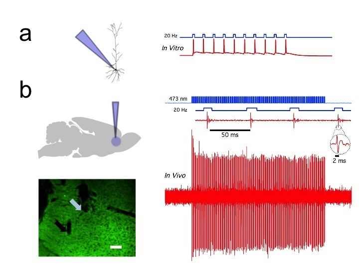
In vivo and in vitro recordings (by the Cooper laboratory) of individual CAMKII AAV-ChR2 expressing pyramidal neurons within the prefrontal cortex demonstrated high fidelity action potential output with short pulses of blue light at 20 Hz (Figure 1). The same group recorded complete green light-induced silencing of spontaneous activity in the same prefrontal cortical neuronal population expressing an AAV-NpHR vector (Figure 2).
Heart
Optogenetics was applied on atrial cardiomyocytes to end spiral wave arrhythmias, found to occur in atrial fibrillation, with light. This method is still in the development stage. A recent study explored the possibilities of optogenetics as a method to correct for arrythmias and resynchronize cardiac pacing. The study introduced Channelrhodopsin-2 into cardiomyocytes in ventricular areas of hearts of transgenic mice and performed in vitro studies of photostimulation on both open-cavity and closed-cavity mice. Photostimulation led to increased activation of cells and thus increased ventricular contractions resulting in increasing heart rates. In addition, this approach has been applied in cardiac resynchronization therapy (CRT) as a new biological pacemaker as a substitute for electrode based-CRT. Lately, optogenetics has been used in the heart to defibrillate ventricular arrhythmias with local epicardial illumination, a generalized whole heart illumination or with customized stimulation patterns based on arrhythmogenic mechanisms in order to lower defibrillation energy.
Spiral ganglion
Optogenetic stimulation of the spiral ganglion in deaf mice restored auditory activity. Optogenetic application onto the cochlear region allows for the stimulation or inhibition of the spiral ganglion cells (SGN). In addition, due to the characteristics of the resting potentials of SGN's, different variants of the protein Channelrhodopsin-2 have been employed such as Chronos and CatCh. Chronos and CatCh variants are particularly useful in that they have less time spent in their deactivated states, which allow for more activity with less bursts of blue light emitted. The result being that the LED producing the light would require less energy and the idea of cochlear prosthetics in association with photo-stimulation, would be more feasible.
Brainstem
Optogenetic stimulation of a modified red-light excitable channelrhodopsin (ReaChR) expressed in the facial motor nucleus enabled minimally invasive activation of motoneurons effective in driving whisker movements in mice. One novel study employed optogenetics on the Dorsal Ralphe Nucleus to both activate and inhibit dopaminergic release onto the Ventral Tegmental Area. To produce activation transgenic mice were infected with Channelrhodopsin-2 with a TH-Cre promoter and to produce inhibition the hyperpolarizing opsin NpHR was added onto the TH-Cre promoter. Results showed that optically activating dopaminergic neurons led to an increase in social interactions, and their inhibition decreased the need to socialize only after a period of isolation.
Precise temporal control of interventions
The currently available optogenetic actuators allow for the accurate temporal control of the required intervention (i.e. inhibition or excitation of the target neurons) with precision routinely going down to the millisecond level. Therefore, experiments can now be devised where the light used for the intervention is triggered by a particular element of behavior (to inhibit the behavior), a particular unconditioned stimulus (to associate something to that stimulus) or a particular oscillatory event in the brain (to inhibit the event). This kind of approach has already been used in several brain regions:
Hippocampus
Sharp waves and ripple complexes (SWRs) are distinct high frequency oscillatory events in the hippocampus thought to play a role in memory formation and consolidation. These events can be readily detected by following the oscillatory cycles of the on-line recorded local field potential. In this way the onset of the event can be used as a trigger signal for a light flash that is guided back into the hippocampus to inhibit neurons specifically during the SWRs and also to optogenetically inhibit the oscillation itself These kinds of "closed-loop" experiments are useful to study SWR complexes and their role in memory.