
Specialty dermatology ICD-9-CM 448.1, 216.0-216.9 | ICD-10 I78.1, D22 MeSH D009506 | |
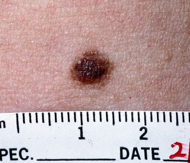 | ||
Nevus (plural: nevi) is a nonspecific medical term for a visible, circumscribed, chronic lesion of the skin or mucosa. The term originates from nævus, which is Latin for "birthmark," however, a nevus can be either congenital (present at birth) or acquired. Common terms, including mole, birthmark, and beauty mark, are used to describe nevi, but these terms do not distinguish specific types of nevi from one another.
Contents
Classification
The term nevus is applied to a number of conditions caused by neoplasias and hyperplasias of melanocytes, as well as a number of pigmentation disorders, both hypermelanotic (containing increased melanin, the pigment responsible for skin color) and hypomelanotic (containing decreased melanin).
Acquired
Congenital
Acquired
Congenital
Additional types of nevi do not involve disorders of pigmentation or melanocytes. These additional nevi represent hamartomatous proliferations of the epithelium, connective tissue, and vascular malformations.
Epidermal nevi
These nevi represent excess growth of specific cells types found in the skin, including those that make up oil and sweat glands.
Connective tissue nevi
These nevi represent abnormalities of collagen in the dermis, the deep layer of the skin.
Vascular nevi
These nevi represent excess growth of blood vessels, including capillaries.
Diagnosis
Nevi are typically diagnosed clinically with the naked eye or using dermatoscopy. More advanced imaging tests are available for distinguishing melanocytic nevi from melanoma, including computerized dermoscopy and image analysis. The management of nevi depends on the type of nevus and the degree of diagnostic uncertainty. Some nevi are known to be benign, and may simply be monitored over time. Others may warrant more thorough examination and biopsy for histopathological examination (looking at a sample of skin under a microscope to detect unique cellular features). For example, a clinician may want to determine whether a pigmented nevus is a type of melanocytic nevus, dysplastic nevus, or melanoma as some of these skin lesions pose a risk for malignancy. The ABCDE criteria (asymmetry, border irregularity, color variegation, diameter > 6 mm, and evolution) are often used to distinguish nevi from melanomas in adults, while modified criteria (amelanosis, bleeding or bumps, uniform color, small diameter or de novo, and evolution) can be used when evaluating suspicious lesions in children. In addition to histopathological examination, some lesions may also warrant additional tests to aid in diagnosis, including special stains, immunohistochemistry, and electron microscopy.
Differential diagnoses
Hypermelanotic nevi must be differentiated from other types of pigmented skin lesions, including:
Management
The management of a nevus depends on the specific diagnosis, however, the options for treatment generally include the following modalities:
Destruction
Surgery
The decision to observe or treat a nevus may depend on a number of factors, including cosmetic concerns, irritative symptoms (e.g., pruritus), ulceration, infection, and concern for potential malignancy.
Syndromes
The term nevus is included in the names of multiple dermatologic syndromes:
Etymology
A nevus may also be spelled naevus. The plural is nevi or naevi. The word is from nævus, Latin for "birthmark".