
Domain Eukaryota | Scientific name Naegleria fowleri Rank Species Higher classification Naegleria | |
 | ||
Similar Naegleria, Amoeba, Balamuthia mandrillaris, Entamoeba, Naegleria gruberi | ||
Naegleria fowleri, colloquially known as the "brain-eating amoeba", is a species of the genus Naegleria, belonging to the phylum Percolozoa. It is a free-living, bacteria-eating amoeba that can be pathogenic, causing a fulminant (sudden and severe) brain infection called naegleriasis, also known as primary amoebic meningoencephalitis (PAM). This microorganism is typically found in bodies of warm freshwater, such as ponds, lakes, rivers, and hot springs. It is also found in the soil near warm-water discharges of industrial plants, and in unchlorinated or minimally-chlorinated swimming pools. It can be seen in either an amoeboid or temporary flagellate stage.
Contents
Life cycle

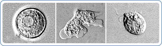
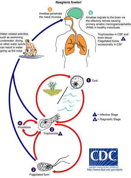
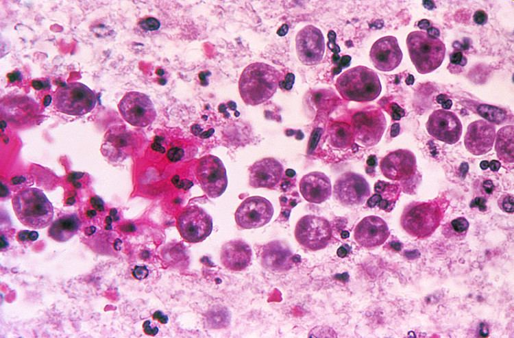
Naegleria fowleri is a thermophilic (heat-loving), free-living amoeba. It is found in warm and hot freshwater ponds, lakes and rivers, and in the very warm water of hot springs. As the water temperature rises, its numbers increase. The amoeba was identified in the 1960s in Australia but appears to have evolved in the United States. N. fowleri occurs in three forms – as a cyst, a trophozoite (ameboid), and a biflagellate (it has two flagella). It does not form a cyst in human tissue, where only the amoeboid trophozoite stage exists. The flagellate form can exist in the cerebrospinal fluid.
Cyst stage
The cyst form is spherical and about 7-15 µm in diameter. They are smooth, and have a single-layered wall with a single nucleus. Cysts are naturally resistant to environmental factors, so as to increase the chances of survival until better conditions occur. Trophozoites encyst due to unfavorable conditions. Factors that induce cyst formation include a lack of food, overcrowding, desiccation, accumulation of waste products, and cold temperatures. When conditions improve, the amoeba can escape through the pore, or ostiole, seen in the middle of the cyst. N. fowleri has been found to encyst at temperatures below 10 °C (50 °F).
Trophozoite stage
The trophozoite is the feeding, dividing, and infective stage for humans. The trophozoite attaches to olfactory epithelium, where it follows the olfactory cell axon through the cribriform plate (in the nasal cavity) to the brain. This reproductive stage of the protozoan organism, which transforms near 25 °C (77 °F) and grows fastest around 42 °C (106.7 °F), proliferates by binary fission. The trophozoites are characterized by a nucleus and a surrounding halo. They travel by pseudopodia, which means that they extend parts of their body's cell membrane (the pseudopods) and then fill them with plasma to force locomotion. The pseudopods form at different points along the cell, thus allowing the trophozoite to change directions. In their free-living state, trophozoites feed on bacteria. In tissues, they phagocytize (consume by enclosing and then digesting prey) red blood cells and white blood cells, destroying tissue.
Flagellate

The flagellate is pear-shaped and biflagellate: this means that it has two flagella. This stage can be inhaled into the nasal cavity during swimming or diving. This biflagellate form occurs when trophozoites are exposed to a change in ionic concentration, such as placement in distilled water. The flagellate form does not exist in human tissue, but can exist in the cerebrospinal fluid. Once inside the nasal cavity, the flagellated form transforms into a trophozoite. The transformation of trophozoite to flagellate occurs within a few hours.
Ecology
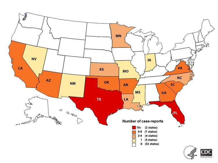
Naegleria fowleri are amoeboflagellates that inhabit soil and water. Naegleria fowleri is more sensitive to drying and pH (acid levels) than other amoeboflagellates. It cannot survive in sea water. This amoeba is able to grow best at moderately elevated temperatures making summer month cases more likely. N. fowleri is thermotolerant and able to survive 45 °C (113 °F). Warm, fresh water with a sufficient supply of bacterial food provide a habitat for amoebae. Man-made bodies of water, disturbed natural habitats, or areas with soil and unchlorinated/unfiltered water are locations where many amoebic infections have happened.
N. fowleri seems to thrive during periods of disturbance; the flagellate-empty hypothesis explains that Naglerias success may be due to decreased competition from decreased amount of the normal, thermosensitive protozoal fauna. In other words, N. fowleri thrives in the absence of other predators consuming its food supply. This hypothesis suggests that human disturbances such as thermal pollution increase N. fowleri abundance by removing their resource competitors. Ameoboflagellates have a motile flagellate stage that is designed for dispersal which is advantageous if an environment has been cleared out of competing organisms.
Pathogenicity
N. fowleri can cause an often lethal infection of the brain called naegleriasis (also known as primary amoebic meningoencephalitis (PAM), amoebic encephalitis/meningitis, or simply Naegleria infection). Infections most often occur when water containing N. fowleri is inhaled through the nose, where it then enters the nasal and olfactory nerve tissue, traveling to the brain through the cribriform plate. N. fowleri normally eat bacteria, but during human infections, the trophozoites consume astrocytes and neurons. The reason why N. fowleri prefers to pass across the cribriform plate has remained unknown, but the neurotransmitter acetylcholine has been suggested to act as a stimulus, as a structural homolog of animal CHRM1 has been shown to present in Naegleria and Acanthamoeba.
It takes up to 5 days (1–12 days average) for symptoms to appear after nasal exposure to N. fowleri flagellates. Initial symptoms may include headache, fever, nausea, or vomiting. Later symptoms can include stiff neck, confusion, lack of attention, loss of balance, seizures, and hallucinations. Once symptoms begin to appear, death will usually occur within two weeks. A person infected with N. fowleri cannot spread the infection to another person.
The core antimicrobial treatment consists of antifungal drug amphotericin B, but the fatality rate even with this treatment is greater than 95%. New treatments are being sought. Miltefosine, an antiparasitic, has been used in a few cases with mixed results.