
Domain Eukaryota Higher classification Acanthamoebidae | Order Centramoebida Scientific name Acanthamoeba Rank Genus | |
 | ||
Similar Naegleria, Balamuthia mandrillaris, Brain‑eating amoeba, Amoeba, Entamoeba | ||
Parasitology acanthamoeba
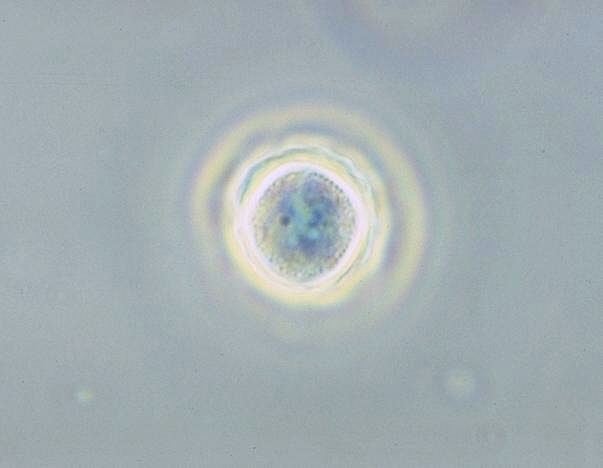
Acanthamoeba is a genus of amoebae, one of the most common single-celled eukaryote in soil, and frequently found in fresh water and other habitats. Acanthamoeba has two evolutive forms, the metabolically active trophozoite and a dormant, stress resistant cyst. Trophozoites are small, usually 15 to 35 μm in length and amoeboid in shape. In nature, Acanthamoeba species are free-living bacterivores, but in certain situations they can cause infections (Acanthamebiasis) in humans and other animals.
Contents
- Parasitology acanthamoeba
- Corneal transplantation for acanthamoeba keratitis
- Distribution
- Role in disease
- Granulomatous amoebic encephalitis
- Acanthamoebic keratitis
- Bacterial infection
- Ecology
- Role as a model organism
- Endosymbionts of Acanthamoeba
- Giant viruses
- Diversity
- References
Corneal transplantation for acanthamoeba keratitis
Distribution
Acanthamoeba spp. are among the most prevalent protozoa found in the environment. They are distributed worldwide, and have been isolated from soil, air, sewage, seawater, chlorinated swimming pools, domestic tap water, bottled water, dental treatment units, hospitals, air-conditioning units, and contact lens cases. Additionally, Acanthamoeba have been isolated from human skin, nasal cavities, throats, and intestines, as well as plants and other mammals.
Role in disease
Diseases caused by Acanthamoeba include keratitis and granulomatous amoebic encephalitis (GAE). The latter is often but not always seen in immunosuppressed patients. GAE is caused by the amoeba entering the body through an open wound then spreading to the brain. The combination of host immune response and amoebal proteases causes massive brain swelling resulting in death in ~95% of those infected.
Granulomatous amoebic encephalitis
Granulomatous amoebic encephalitis is caused by amoeba infection of the central nervous system. It is characterized by neurological symptoms including headache, seizures, and mental status abnormalities. These worsen progressively over weeks to months, leading to death in most patients. Infection is generally associated with underlying conditions such as immunodeficiency, diabetes, malignancies, malnutrition, systemic lupus erythematosus, and alcoholism. The parasite enters the body through cuts in the skin or by being inhaled into the upper respiratory tract. The parasite then spreads through the blood into the central nervous system. Acanthamoeba crosses the blood–brain barrier by means that are not yet understood. Subsequent invasion of the connective tissue and induction of pro-inflammatory responses leads to neuronal damage that can be fatal within days. Pure granulomatous lesions are rare in patients with AIDS and other related immunodeficiency states as the patients do not have adequate numbers of CD+ve T-cells to mount a granulomatous response to Acanthamoeba infection in CNS and other organ and tissues. A perivascular cuffing with amoebae in necrotic tissue is usual finding in the AIDS and related T-cell immunodeficiency conditions. A post mortem biopsy reveals severe oedema and hemorrhagic necrosis. A patient who has contracted this illness usually displays subacute symptoms, including altered mental status, headaches, fever, neck stiffness, seizures, focal neurological signs (such as cranial nerve palsies and coma), all leading to death within one week to several months. Due to the rarity of this parasite and a lack of knowledge, there are currently no good diagnoses or treatments for Acanthamoeba infection. Acanthamoeba keratitis cases in the past, when were managed by atropine given as an adjuvant therapy without anti-parasitic drugs added to the regime, had been reported to halt the vision loss. Recent publications debate these successes related to an action of Atropine on muscarinic receptors on Acanthamoeba spp.
Infection usually mimics that of bacterial leptomeningitis, tuberculous meningitis, or viral encephalitis. The misdiagnosis often leads to erroneous, ineffective treatment. In the case that the Acanthamoeba is diagnosed correctly, the current treatments, such as amphotericin B, rifampicin, trimethoprim-sulfamethoxazole, ketoconazole, fluconazole, sulfadiazine, or albendazole, are only tentatively successful. Correct and timely diagnosis, as well as improved treatment methods and an understanding of the parasite are important factors in improving the outcome of infection by Acanthamoeba. A recent paper published in 2013 has shown substantial effects of some FDA approved drugs with a kill rate above 90%. These results were in-vitro effects, but as the drugs are already approved, human infections can be targeted after dose calculations in clinical trials with them.
Acanthamoebic keratitis
When present in the eye, Acanthamoeba strains can cause acanthamoebic keratitis, which may lead to corneal ulcers or even blindness. This condition occurs most often among contact lens wearers who do not properly disinfect their lenses, exacerbated by a failure to wash hands prior to handling the lenses. Multipurpose contact lens solutions are largely ineffective against Acanthamoeba, whereas hydrogen peroxide-based solutions have good disinfection characteristics.
In May 2007, Advanced Medical Optics, manufacturer of Complete Moisture Plus Contact Lens Solution products, issued a voluntary recall of their Complete Moisture Plus solutions. The fear was that contact lens wearers who used their solution were at higher risk of acanthamoebic keratitis than contact lens wearers who used other solutions. The manufacturer recalled the product after the Centers for Disease Control in the United States found that 21 individuals had possibly received an Acanthamoeba infection after using Complete Moisture Plus in the month prior to diagnosis.
Bacterial infection
Several species of bacteria which can cause human disease are also able to infect and replicate within Acanthamoeba species. These include Legionella pneumophila, Pseudomonas aeruginosa, and some strains of Escherichia coli and Staphylococcus aureus. For some of these bacteria, replication inside Acanthamoeba has been associated with enhanced growth in macrophages, and increased resistance to some antibiotics. Furthermore, due to the high prevalence of Acanthamoeba in the environment, it has been proposed that these amoeba could serve as an environmental reservoir for some human pathogens.
Ecology
A. castellanii can be found at high densities in various soil ecosystems. It preys on bacteria, but also fungi and other protozoa.
This species is able to lyse bacteria and produce a wide range of enzymes, such as cellulases or chitinases, and probably contributes to the breakdown of organic matter in soil, contributing to the microbial loop.
Role as a model organism
Because Acanthamoeba does not differ greatly at the ultrastructural-level from a mammalian cell, it is an attractive model for cell biology studies. Acanthamoeba is important in cellular microbiology, environmental biology, physiology, cellular interactions, molecular biology, biochemistry, and evolutionary studies, due to the organisms' versatile roles in the ecosystem and ability to capture prey by phagocytosis, act as vectors and reservoirs for microbial pathogens, and to produce serious human infections. In addition, Acanthamoeba has been used extensively to understand the molecular biology of cell motility and cancer cell dormancy by in-depth exploration of the process of encystation
The recently available Acanthamoeba genome sequence revealed several orthologs of genes employed in meiosis of sexual eukaryotes. These genes included Spo11, Mre11, Rad50, Rad51, Rad52, Mnd1, Dmc1, Msh and Mlh. This finding suggests that Acanthamoeba is capable of some form of meiosis and may be able to undergo sexual reproduction. Furthermore, since Acanthamoeba diverged early from the eukaryotic family tree, these results suggest that meiosis was present early in eukaryotic evolution.
Owing to its ease and economy of cultivation, the Neff strain of A. castellanii discovered in a pond in Golden Gate Park in the 1960s, has been effectively used as a classic model organism in the field of cell biology. From just 30 liters of simple medium inoculated with A. castellanii, approximately one kilogram of cells can be obtained after several days of aerated culture at room temperature. Pioneered in the laboratory of Edward D. Korn at the National Institutes of Health (NIH), many important biological molecules have been discovered and their pathways elucidated using the Acanthamoeba model. Thomas Dean Pollard applied this model at the NIH, Harvard Medical School, Johns Hopkins University School of Medicine, and the Salk Institute for Biological Studies to discover and characterize many proteins that are essential for cell motility, not only in amoebas, but also in many other eukaryotic cells, especially those of the human nervous and immune systems, the developing embryo, and cancer cells. Acanthamoeba also has served as a model to study the evolution of certain G-Proteins. This unicellular eukaryote expresses few GPCRs over its cell membrane that serve vital role for the microorganism, structural homology bioinformatics tools have been used to show the presence of a homolog of human M1-Muscarinic receptor in Acanthamoeba castellanii. Blocking these muscarinic receptors in past studies has proven to be amoebicidal in Acanthamoeba spp.
Endosymbionts of Acanthamoeba
Acanthamoeba spp. contain diverse bacterial endosymbionts that are similar to human pathogens, so they are considered to be potential emerging human pathogens. The exact nature of these symbionts and the benefit they represent for the amoebal host still have to be clarified.
These include Legionella and Legionella-like pathogens.
Giant viruses
The giant viruses Mimivirus, Megavirus and Pandoravirus infect Acanthamoeba.
Currently Acanthamoeba is the only species that is a host for such huge viruses (who have more than 1000 protein-coding genes; for instance, Pandoravirus has about 2500 protein-coding genes in its genome).
Diversity
Acanthamoeba can be distinguished from other genera of amoeba based on morphological characteristics. However, differentiating one species of Acanthamoeba from another by morphology has proven difficult. Based on 18S rDNA sequencing, known Acanthamoeba strains can be organized into 12 groups, denoted T1-T12. Most disease-causing isolates belong to type T4.
Below is a list of described species of Acanthamoeba, with sequence types noted where known. Species which have been identified in diseased patients are marked with *.