
Entrez 2660 | Ensembl ENSG00000138379 | |
 | ||
Aliases MSTN, GDF8, MSLHP, myostatin External IDs OMIM: 601788 MGI: 95691 HomoloGene: 3850 GeneCards: MSTN | ||
Myostatin (also known as growth differentiation factor 8, abbreviated GDF-8) is a myokine, a protein produced and released by myocytes that acts on muscle cells' autocrine function to inhibit myogenesis: muscle cell growth and differentiation. In humans it is encoded by the MSTN gene. Myostatin is a secreted growth differentiation factor that is a member of the TGF beta protein family.
Contents
- Discovery and sequencing
- Structure and mechanism of action
- Double muscled cattle
- Whippets
- Rabbits and Goats
- Heart
- Mutations
- In humans
- Therapeutic potential
- Gene doping
- Television
- References
Animals either lacking myostatin or treated with substances that block the activity of myostatin have significantly more muscle mass. Furthermore, individuals who have mutations in both copies of the myostatin gene have significantly more muscle mass and are stronger than normal. There is hope that studies into myostatin may have therapeutic application in treating muscle wasting diseases such as muscular dystrophy.
Discovery and sequencing
The gene encoding myostatin was discovered in 1997 by geneticists Se-Jin Lee and Alexandra McPherron who produced a knockout strain of mice that lack the gene, and have approximately twice as much muscle as normal mice. These mice were subsequently named "mighty mice".
Naturally occurring deficiencies of myostatin of various sorts have been identified in some breeds of cattle, sheep, whippets, and humans. In each case the result is a dramatic increase in muscle mass.
Structure and mechanism of action
Human myostatin consists of two identical subunits, each consisting of 109 (NCBI database claims human myostatin is 375 residues long) amino acid residues [note the full length gene encodes a 375AA prepro-protein which is proteolytically processed to its shorter active form]. Its total molecular weight is 25.0 kDa. The protein is inactive until a protease cleaves the NH2-terminal, or "pro-domain" portion of the molecule, resulting in the active COOH-terminal dimer. Myostatin binds to the activin type II receptor, resulting in a recruitment of either coreceptor Alk-3 or Alk-4. This coreceptor then initiates a cell signaling cascade in the muscle, which includes the activation of transcription factors in the SMAD family - SMAD2 and SMAD3. These factors then induce myostatin-specific gene regulation. When applied to myoblasts, myostatin inhibits their differentiation into mature muscle fibers.
Myostatin also inhibits Akt, a kinase that is sufficient to cause muscle hypertrophy, in part through the activation of protein synthesis. However, Akt is not responsible for all of the observed muscle hyperthrophic effects which are mediated by myostatin inhibition Thus myostatin acts in two ways: by inhibiting muscle differentiation, and by inhibiting Akt-induced protein synthesis.
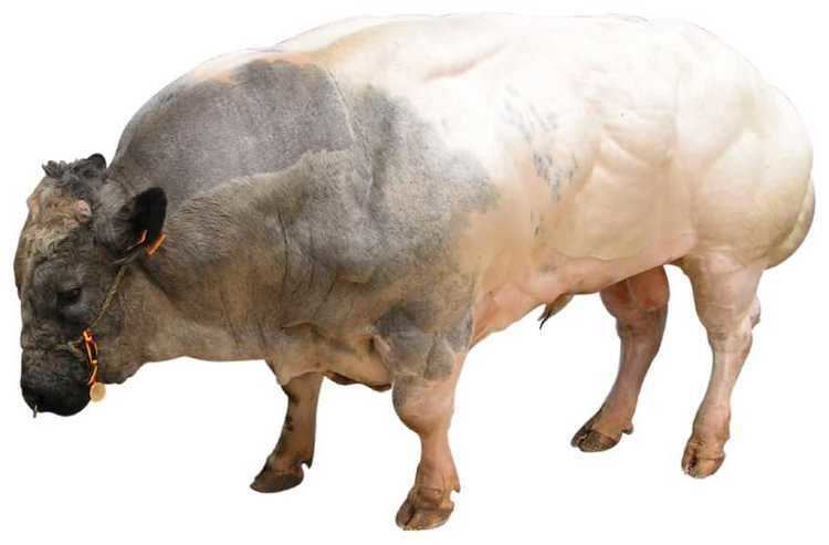
Double muscled cattle
After that discovery, several laboratories cloned and established the nucleotide sequence of a myostatin gene in two breeds of cattle, Belgian Blue and Piedmontese. They found mutations in the myostatin gene (various mutations in each breed) which in one way or another lead to absence of functional myostatin. Unlike mice with a damaged myostatin gene, in these cattle breeds the muscle cells multiply rather than enlarge. People describe these cattle breeds as "double muscled", but the total increase in all muscles is no more than 40%.
Animals lacking myostatin or animals treated with substances such as follistatin that block the binding of myostatin to its receptor have significantly larger muscles. Thus, reduction of myostatin could potentially benefit the livestock industry, with even a 20 percent reduction in myostatin levels potentially having a large effect on the development of muscles.
However, the animal breeds developed as homozygous for myostatin deficiency have reproduction issues due to their unusually heavy and bulky offspring, and require special care and a more expensive diet to achieve a superior yield. This negatively affects economics of myostatin-deficient breeds to the point where they do not usually offer an obvious advantage. While hypertrophic meat (e.g. from Piedmontese beef) has a place on the specialist market due to its unusual properties, at least for purebred myostatin-deficient strains the expenses and (especially in cattle) necessity of veterinary supervision place them at a disadvantage in the bulk market.
Whippets
Whippets can have a mutation of the myostatin which involves a 2 base pair deletion, and results in a truncated, and likely inactive, myostatin protein.
Animals with a homozygous deletion have an unusual body shape, with a broader head, pronounced overbite, shorter legs, and thicker tails and are called "bully whippets" by the breeding community. Although significantly more muscular, they are less able runners than other whippets. On the other hand, whippets that were heterozygous for the mutation were significantly over-represented in the top racing classes.
Rabbits and Goats
In 2016, the CRISPR/Cas9 system was used to genetically engineer rabbits and goats with no functional copies of the myostatin gene. In both cases the resulting animals were significantly more muscular. However, rabbits without myostatin also exhibited an enlarged tongue, a higher rate of still births, and a reduced lifespan.
Heart
Myostatin is expressed at very low levels in cardiac myocytes. Although its presence has been noted in cardiomyocytes of both fetal and adult mice, its physiological function remains uncertain. However, it has been suggested that fetal cardiac myostatin may play a role in early heart development.
Myostatin is produced as promyostatin, a precursor protein kept inactive by the latent TGF-β binding protein 3 (LTBP3). Pathological cardiac stress promotes N-terminal cleavage by furin convertase to create a biologically active C-terminal fragment. The mature myostatin is then segregated from the latent complex via proteolytic cleavage by BMP-1 and tolloid metallopreoteinases. Free myostatin is able to bind its receptor, ActRIIB, and increase SMAD2/3 phosphorylation. The latter produces a heteromeric complex with SMAD4, inducing myostatin translocation into the cardiomyocyte nucleus to modulate transcription factor activity. Manipulating the muscle creatinine kinase promoter can modulate myostatin expression, although it has only been observed in male mice thus far.
Myostatin may inhibit cardiomyocyte proliferation and differentiation by manipulating cell cycle progression. This argument is supported by the fact that myostatin mRNA is poorly expressed in proliferating fetal cardiomyocytes. In vitro studies indicate that myostatin promotes SMAD2 phosphorylation to inhibit cardiomyocyte proliferation. Furthermore, myostatin has been shown to directly prevent cell cycle G1 to S phase transition by decreasing levels of cyclin-dependent kinase complex 2 (CDK2) and by increasing p21 levels.
Growth of cardiomyocytes may also be hindered by myostatin-regulated inhibition of protein kinase p38 and the serine-threonine protein kinase Akt, which typically promote cardiomyocyte hypertrophy. However, increased myostatin activity only occurs in response to specific stimuli, such as in pressure stress models, in which cardiac myostatin induces whole-body muscular atrophy.
Physiologically, minimal amounts of cardiac myostatin are secreted from the myocardium into serum, having a limited effect on muscle growth. However, increases in cardiac myostatin can increase its serum concentration, which may cause skeletal muscle atrophy. Pathological states that increase cardiac stress and promote heart failure can induce a rise in both cardiac myostatin mRNA and protein levels within the heart. In ischemic or dilated cardiomyopathy, increased levels of myostatin mRNA have been detected within the left ventricle.
As a member of the TGF-β family, myostatin may play a role in post-infarct recovery. It has been hypothesized that hypertrophy of the heart induces an increase in myostatin as a negative feedback mechanism in an attempt to limit further myocyte growth. This process includes mitogen-activated protein kinases and binding of the MEF2 transcription factor within the promoter region of the myostatin gene. Increases in myostatin levels during chronic heart failure have been shown to cause cardiac cachexia. Systemic inhibition of cardiac myostatin with the JA-16 antibody maintains overall muscle weight in experimental models with pre-existing heart failure.
Myostatin also alters excitation-contraction (EC) coupling within the heart. A reduction in cardiac myostatin induces eccentric hypertrophy of the heart, and increases its sensitivity to beta-adrenergic stimuli by enhancing Ca2+ release from the SR during EC coupling. Also, phospholamban phosphorylation is increased in myostatin-knockout mice, leading to an increase in Ca2+ release into the cytosol during systole. Therefore, minimizing cardiac myostatin may improve cardiac output.
Mutations
A technique for detecting mutations in myostatin variants has been developed. Mutations that reduce the production of functional myostatin lead to an overgrowth of muscle tissue. Myostatin-related muscle hypertrophy has an incomplete autosomal dominance pattern of inheritance. People with a mutation in both copies of the MSTN gene in each cell (homozygotes) have significantly increased muscle mass and strength. People with a mutation in one copy of the MSTN gene in each cell (heterozygotes) have increased muscle bulk, but to a lesser degree.
In humans
In 2004, a German boy was diagnosed with a mutation in both copies of the myostatin-producing gene, making him considerably stronger than his peers. His mother has a mutation in one copy of the gene.
An American boy born in 2005 was diagnosed with a clinically similar condition but with a somewhat different cause: his body produces a normal level of functional myostatin; but, because he is stronger and more muscular than most others his age, it is believed that a defect in his myostatin receptors prevents his muscle cells from responding normally to myostatin. He appeared on the television show, World's Strongest Toddler.
Therapeutic potential
Further research into myostatin and the myostatin gene may lead to therapies for muscular dystrophy. The idea is to introduce substances that block myostatin. A monoclonal antibody specific to myostatin increases muscle mass in mice and monkeys.
A two-week treatment of normal mice with soluble activin type IIB receptor, a molecule that is normally attached to cells and binds to myostatin, leads to a significantly increased muscle mass (up to 60%). It is thought that binding of myostatin to the soluble activin receptor prevents it from interacting with the cell-bound receptors.
It remains unclear as to whether long-term treatment of muscular dystrophy with myostatin inhibitors is beneficial, as the depletion of muscle stem cells could worsen the disease later on. As of 2012, no myostatin-inhibiting drugs for humans are on the market. An antibody genetically engineered to neutralize myostatin, stamulumab, which was under development by pharmaceutical company Wyeth, is no longer under development. Some athletes, eager to get their hands on such drugs, turn to the internet where fake "myostatin blockers" are being sold.
Myostatin levels are effectively decreased by creatine supplementation.
Myostatin levels can be temporarily reduced using a cholesterol-conjugated siRNA gene knockdown.
Gene doping
See Gene doping
Inhibition of myostatin leads to muscle hyperplasia and hypertrophy. Myostatin inhibitors can improve athletic performance and therefore there is a concern these inhibitors might be abused in the field of sports. However, studies in mice suggest that myostatin inhibition does not directly increase the strength of individual muscle fibers.
Television
In The Incredible Hulk episode "Death In The Family" a nurse gives a patient a dose of myostatin, but Dr. David Banner recognizes that it is not true myostatin because the liquid is brown not clear.