
Group Group V ((−)ssRNA) | ||
 | ||
Marburg virus is a hemorrhagic fever virus of the Filoviridae family of viruses and a member of the species Marburg marburgvirus, genus Marburgvirus. Marburg virus (MARV) causes Marburg virus disease in humans and nonhuman primates, a form of viral hemorrhagic fever. The virus is considered to be extremely dangerous. The WHO rates it as a Risk Group 4 Pathogen (requiring biosafety level 4-equivalent containment). In the United States, the NIH/National Institute of Allergy and Infectious Diseases ranks it as a Category A Priority Pathogen and the Centers for Disease Control and Prevention lists it as a Category A Bioterrorism Agent. It is also listed as a biological agent for export control by the Australia Group.
Contents
- Discovery
- Nomenclature
- Human disease
- Genome
- Structure
- Entry
- Replication
- Ecology
- Evolution
- Biological weapon
- References
In 2009, expanded clinical trials of an Ebola and Marburg vaccine began in Kampala, Uganda.
Discovery
Marburg virus was first described in 1967. It was noticed during small outbreaks in the German cities Marburg and Frankfurt and the Yugoslav capital Belgrade in the 1960s. German workers were exposed to tissues of infected grivet monkeys (Chlorocebus aethiops) at the city's former main industrial plant, the Behringwerke, then part of Hoechst, and today of CSL Behring. During these outbreaks, 31 people became infected and seven of them died. MARV is a Select Agent.
Nomenclature
The virus is one of two members of the species Marburg marburgvirus, which is included in the genus Marburgvirus, family Filoviridae, order Mononegavirales. The name Marburg virus is derived from Marburg (the city in Hesse, Germany, where the virus was first discovered) and the taxonomic suffix virus.
According to the rules for taxon naming established by the International Committee on Taxonomy of Viruses (ICTV), the name Marburg virus is always to be capitalized, but is never italicized, and may be abbreviated (with MARV being the official abbreviation).
Marburg virus was first introduced under this name in 1967. In 2005, the virus name was changed to Lake Victoria marburgvirus, which unfortunately was the same spelling as its species Lake Victoria marburgvirus. However, most scientific articles continued to refer to Marburg virus. Consequently, in 2010, the name Marburg virus was reinstated and the species name changed. A previous abbreviation for the virus was MBGV.
Human disease
MARV is one of two Marburg viruses that causes Marburg virus disease (MVD) in humans (in the literature also often referred to as Marburg hemorrhagic fever, MHF). The other one is Ravn virus (RAVV). Both viruses fulfill the criteria for being a member of the species Marburg marburgvirus because their genomes diverge from the prototype Marburg marburgvirus or the Marburg virus variant Musoke (MARV/Mus) by <10% at the nucleotide level.
Genome
Like all mononegaviruses, marburgvirions contain non-infectious, linear nonsegmented, single-stranded RNA genomes of negative polarity that possess inverse-complementary 3' and 5' termini, do not possess a 5' cap, are not polyadenylated, and are not covalently linked to a protein. Marburgvirus genomes are approximately 19 kb long and contain seven genes in the order 3'-UTR-NP-VP35-VP40-GP-VP30-VP24-L-5'-UTR. The genomes of the two different marburgviruses (MARV and RAVV) differ in sequence.
Structure
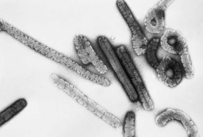
Like all filoviruses, marburgvirions are filamentous particles that may appear in the shape of a shepherd's crook or in the shape of a "U" or a "6", and they may be coiled, toroid, or branched. Marburgvirions are generally 80 nm in width, but vary somewhat in length. In general, the median particle length of marburgviruses ranges from 795 to 828 nm (in contrast to ebolavirions, whose median particle length was measured to be 974–1,086 nm ), but particles as long as 14,000 nm have been detected in tissue culture. Marburgvirions consist of seven structural proteins. At the center is the helical ribonucleocapsid, which consists of the genomic RNA wrapped around a polymer of nucleoproteins (NP). Associated with the ribonucleoprotein is the RNA-dependent RNA polymerase (L) with the polymerase cofactor (VP35) and a transcription activator (VP30). The ribonucleoprotein is embedded in a matrix, formed by the major (VP40) and minor (VP24) matrix proteins. These particles are surrounded by a lipid membrane derived from the host cell membrane. The membrane anchors a glycoprotein (GP1,2) that projects 7 to 10 nm spikes away from its surface. While nearly identical to ebolavirions in structure, marburgvirions are antigenically distinct.
Entry
Niemann–Pick C1 (NPC1) cholesterol transporter protein appears to be essential for infection with both Ebola and Marburg virus. Two independent studies reported in the same issue of Nature showed that Ebola virus cell entry and replication requires NPC1. When cells from patients lacking NPC1 were exposed to Ebola virus in the laboratory, the cells survived and appeared immune to the virus, further indicating that Ebola relies on NPC1 to enter cells. This might imply that genetic mutations in the NPC1 gene in humans could make some people resistant to one of the deadliest known viruses affecting humans. The same studies described similar results with Marburg virus, showing that it also needs NPC1 to enter cells. Furthermore, NPC1 was shown to be critical to filovirus entry because it mediates infection by binding directly to the viral envelope glycoprotein and that the second lysosomal domain of NPC1 mediates this binding.
In one of the original studies, a small molecule was shown to inhibit Ebola virus infection by preventing the virus glycoprotein from binding to NPC1. In the other study, mice that were heterozygous for NPC1 were shown to be protected from lethal challenge with mouse-adapted Ebola virus. Together, these studies suggest NPC1 may be potential therapeutic target for an Ebola antiviral drug.
Replication
The marburg virus life cycle begins with virion attachment to specific cell-surface receptors, followed by fusion of the virion envelope with cellular membranes and the concomitant release of the virus nucleocapsid into the cytosol. The virus RdRp partially uncoats the nucleocapsid and transcribes the genes into positive-stranded mRNAs, which are then translated into structural and nonstructural proteins. Marburgvirus L binds to a single promoter located at the 3' end of the genome. Transcription either terminates after a gene or continues to the next gene downstream. This means that genes close to the 3' end of the genome are transcribed in the greatest abundance, whereas those toward the 5' end are least likely to be transcribed. The gene order is therefore a simple but effective form of transcriptional regulation. The most abundant protein produced is the nucleoprotein, whose concentration in the cell determines when L switches from gene transcription to genome replication. Replication results in full-length, positive-stranded antigenomes that are in turn transcribed into negative-stranded virus progeny genome copies. Newly synthesized structural proteins and genomes self-assemble and accumulate near the inside of the cell membrane. Virions bud off from the cell, gaining their envelopes from the cellular membrane they bud from. The mature progeny particles then infect other cells to repeat the cycle.
Ecology
In 2009, the successful isolation of infectious MARV was reported from caught healthy Egyptian rousettes (Rousettus aegyptiacus). This isolation, together with the isolation of infectious RAVV, strongly suggests that Old World fruit bats are involved in the natural maintenance of marburgviruses. Further studies are necessary to establish whether Egyptian rousettes are the actual hosts of MARV and RAVV or whether they get infected via contact with another animal and therefore serve only as intermediate hosts. Recently the first experimental infection study of Rousettus aegyptiacus with MARV provided further insight into the possible involvement of these bats in MARV ecology. Experimentally infected bats developed relatively low viremia lasting at least 5 days, but remained healthy and didn't develop any notable gross pathology. The virus also replicated to high titers in major organs (liver and spleen), and organs that might possibly be involved in virus transmission (lung, intestine, reproductive organ, salivary gland, kidney, bladder and mammary gland). The relatively long period of viremia noted in this experiment could possibly also facilitate mechanical transmission by blood sucking arthropods or infection of susceptible vertebrate hosts by direct contact with infected blood.
Evolution
The viral strains fall into two clades - Ravn virus and Marburg virus. The Marburg strains can be divided into two - A and B. The A strains were isolated from Uganda (five from 1967), Kenya (1980) and Angola (2004-2005) while the B strains were from the Democratic Republic of the Congo epidemic (1999-2000) and a group of Ugandan isolates isolated in 2007-2009.
The mean evolutionary rate of the whole genome was 3.3 × 10⁻⁴ substitutions/site/year (credibility interval 2.0-4.8).
The Marburg strains had a mean root time of the most recent common ancestor of 177.9 years ago (95% highest posterior density 87-284) suggesting an origin in the mid 1800s. In contrast the Ravn strains origin dated back to a mean 33.8 years ago (early 1980s). The most probable location of the Marburg virus ancestor was Uganda whereas that of the RAVV ancestor was Kenya.
Biological weapon
The Soviet Union had an extensive offensive and defensive biological weapons program that included MARV. At least three Soviet research institutes had MARV research programs during the Cold War: the Virology Center of the Scientific-Research Institute for Microbiology in Zagorsk (today Sergiev Posad), the Scientific-Production Association "Vektor" (today the State Research Center of Virology and Biotechnology "Vektor") in Koltsovo, and the Irkutsk Scientific-Research Anti-Plague Institute of Siberia and the Far East in Irkutsk. As most performed research was highly classified, it remains unclear how successful the MARV program was. However, Soviet defector Ken Alibek claimed that a weapon filled with MARV was tested at the Stepnogorsk Scientific Experimental and Production Base in Stepnogorsk, Kazakh Soviet Socialist Republic (today Kazakhstan), suggesting that the development of a MARV biological weapon had reached advanced stages. Independent confirmation for this claim is lacking. At least one laboratory accident with MARV, resulting in the death of Koltsovo researcher Nikolai Ustinov, occurred during the Cold War in the Soviet Union and was first described in detail by Alibek.