
Entrez 4537 | Ensembl ENSG00000198840 | |
 | ||
Aliases ND3, MTMT-NADH dehydrogenase, subunit 3 (complex I) External IDs MGI: 102499 HomoloGene: 5018 GeneCards: ND3 | ||
Mitochondrially encoded NADH dehydrogenase 3 is a protein that in humans is encoded by the mitochondrial gene MT-ND3. The ND3 protein is a subunit of NADH dehydrogenase (ubiquinone), which is located in the mitochondrial inner membrane and is the largest of the five complexes of the electron transport chain. Variants of MT-ND3 are associated with Mitochondrial encephalomyopathy, lactic acidosis, and stroke-like episodes (MELAS), Leigh's syndrome (LS) and Leber's hereditary optic neuropathy (LHON).
Contents
Structure
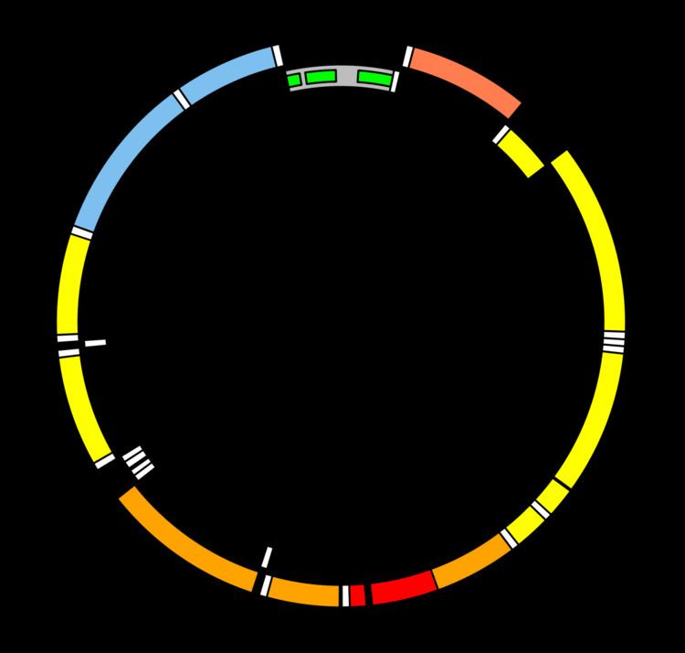
MT-ND3 is located in human mitochondrial DNA from base pair 10,059 to 10,404. The MT-ND3 gene produces a 13 kDa protein composed of 115 amino acids. MT-ND3 is one of seven mitochondrially-encoded subunits of the enzyme NADH dehydrogenase (ubiquinone). Also known as Complex I, it is the largest of the respiratory complexes. The structure is L-shaped with a long, hydrophobic transmembrane domain and a hydrophilic domain for the peripheral arm that includes all the known redox centres and the NADH binding site. MT-ND3 and the rest of the mitochondrially encoded subunits are the most hydrophobic of the subunits of Complex I and form the core of the transmembrane region.
Function
MT-ND3 is a subunit of the respiratory chain Complex I that is believed to belong to the minimal assembly of core proteins required to catalyze NADH dehydrogenation and electron transfer to ubiquinone (coenzyme Q10). Initially, NADH binds to Complex I and transfers two electrons to the isoalloxazine ring of the flavin mononucleotide (FMN) prosthetic arm to form FMNH2. The electrons are transferred through a series of iron-sulfur (Fe-S) clusters in the prosthetic arm and finally to coenzyme Q10 (CoQ), which is reduced to ubiquinol (CoQH2). The flow of electrons changes the redox state of the protein, resulting in a conformational change and pK shift of the ionizable side chain, which pumps four hydrogen ions out of the mitochondrial matrix.
Untranslated extra nucleotide
In the MT-ND3 gene from many species of birds and turtles there is an extra nucleotide that is not translated to protein. Translational frameshifting or RNA editing are alternative explanations for maintenance of the functionality of the ND3 reading frame in birds possessing the one-nucleotide insertion. This extra nucleotide feature suggests that turtles might be related to Archosauria, as evidenced by molecular phylogeny studies. The absence of the extra nucleotide in crocodilians and some birds and turtles might also indicate that the corresponding taxa have lost this feature.
Clinical significance
Pathogenic variants of the mitochondrial gene MT-ND3 are known to cause mtDNA-associated Leigh syndrome, as are variants of MT-ATP6, MT-TL1, MT-TK, MT-TW, MT-TV, MT-ND1, MT-ND2, MT-ND4, MT-ND5, MT-ND6 and MT-CO3. Abnormalities in mitochondrial energy generation result in neurodegenerative disorders like Leigh syndrome, which is characterized by an onset of symptoms between 12 months and three years of age. The symptoms frequently present themselves following a viral infection and include movement disorders and peripheral neuropathy, as well as hypotonia, spasticity and cerebellar ataxia. Roughly half of affected patients die of respiratory or cardiac failure by the age of three. Leigh syndrome is a maternally inherited disorder and its diagnosis is established through genetic testing of the aforementioned mitochondrial genes, including MT-ND3. These complex I genes have been associated with a variety of neurodegenerative disorders, including Leber's hereditary optic neuropathy (LHON), mitochondrial encephalomyopathy with stroke-like episodes (MELAS) and the previously mentioned Leigh syndrome.