
Insertion extensor expansion | ||
 | ||
Artery Latin musculi lumbricales manus | ||
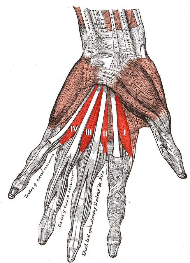
The lumbricals are intrinsic muscles of the hand that flex the metacarpophalangeal joints and extend the interphalangeal joints.
Contents
Structure
There are four of these small, worm-like muscles on each hand. These muscles are unusual in that they do not attach to bone. Instead, they attach proximally to the tendons of flexor digitorum profundus and distally to the extensor expansions.
Innervation
The first and second lumbricals (the most radial two) are innervated by the median nerve. The third and fourth lumbricals (most ulnar two) are innervated by the deep branch of the ulnar nerve.
This is the usual innervation of the lumbricals (occurring in 60% of individuals). However 1:3 (median:ulnar - 20% of individuals) and 3:1 (median:ulnar - 20% of individuals) also exist. The lumbrical innervation always follows the innervation pattern of the associated muscle unit of flexor digitorum profundus (i.e. if the muscle units supplying the tendon to the middle finger are innervated by the median nerve, the second lumbrical will also be innervated by the median nerve).
Blood supply
There are four separate sources of blood supply for these muscles: the superficial palmar arch, the common palmar digital artery, the deep palmar arch, and the dorsal digital artery.
Actions
The lumbrical muscles, with the help of the interosseous muscles, simultaneously flex the metacarpophalangeal joints while extending both interphalangeal joints of the digit on which it inserts. The lumbricals are used during an upstroke in writing.
Other lumbricals
There are also lumbrical muscles of the foot that have a similar action, though these are of less clinical concern.