
ICD-9-CM 161 MeSH D007822 | ICD-10 C32 | |
 | ||
Laryngeal cancer, also known as cancer of the larynx or laryngeal carcinoma, are mostly squamous cell carcinomas, reflecting their origin from the skin of the larynx.
Contents
Cancer can develop in any part of the larynx, but the cure rate is affected by the location of the tumour. For the purposes of tumour staging, the larynx is divided into three anatomical regions: the glottis (true vocal cords, anterior and posterior commissures); the supraglottis (epiglottis, arytenoids and aryepiglottic folds, and false cords); and the subglottis.
Most laryngeal cancers originate in the glottis. Supraglottic cancers are less common, and subglottic tumours are least frequent.
Laryngeal cancer may spread by direct extension to adjacent structures, by metastasis to regional cervical lymph nodes, or more distantly, through the blood stream. Distant metastases to the lung are most common. In 2013 it resulted in 88,000 deaths up from 76,000 deaths in 1990. Five year survival rates in the United States are 60%.
Signs and symptoms
The symptoms of laryngeal cancer depend on the size and location of the tumour. Symptoms may include the following:
Treatment effects can include post-operative changes in appearance, difficulty eating, or loss of voice that may require learning alternate methods of speaking.
Risk factors
Smoking is the most important risk factor for laryngeal cancer. Death from laryngeal cancer is 20 times more likely for heaviest smokers than for nonsmokers. Heavy chronic consumption of alcohol, particularly alcoholic spirits, is also significant. When combined, these two factors appear to have a synergistic effect. Some other quoted risk factors are likely, in part, to be related to prolonged alcohol and tobacco consumption. These include low socioeconomic status, male sex, and age greater than 55 years.
People with a history of head and neck cancer are known to be at higher risk (about 25%) of developing a second cancer of the head, neck, or lung. This is mainly because in a significant proportion of these patients, the aerodigestive tract and lung epithelium have been exposed chronically to the carcinogenic effects of alcohol and tobacco. In this situation, a field change effect may occur, where the epithelial tissues start to become diffusely dysplastic with a reduced threshold for malignant change. This risk may be reduced by quitting alcohol and tobacco.
Diagnosis
Diagnosis is made by the doctor on the basis of a medical history, physical examination, and special investigations which may include a chest x-ray, CT or MRI scans, and tissue biopsy. The examination of the larynx requires some expertise, which may require specialist referral.
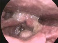
The physical exam includes a systematic examination of the whole patient to assess general health and to look for signs of associated conditions and metastatic disease. The neck and supraclavicular fossa are palpated to feel for cervical adenopathy, other masses, and laryngeal crepitus. The oral cavity and oropharynx are examined under direct vision. The larynx may be examined by indirect laryngoscopy using a small angled mirror with a long handle (akin to a dentist's mirror) and a strong light. Indirect laryngoscopy can be highly effective, but requires skill and practice for consistent results. For this reason, many specialist clinics now use fibre-optic nasal endoscopy where a thin and flexible endoscope, inserted through the nostril, is used to clearly visualise the entire pharynx and larynx. Nasal endoscopy is a quick and easy procedure performed in clinic. Local anaesthetic spray may be used.
If there is a suspicion of cancer, biopsy is performed, usually under general anaesthetic. This provides histological proof of cancer type and grade. If the lesion appears to be small and well localised, the surgeon may undertake excision biopsy, where an attempt is made to completely remove the tumour at the time of first biopsy. In this situation, the pathologist will not only be able to confirm the diagnosis, but can also comment on the completeness of excision, i.e., whether the tumour has been completely removed. A full endoscopic examination of the larynx, trachea, and esophagus is often performed at the time of biopsy.
For small glottic tumours further imaging may be unnecessary. In most cases, tumour staging is completed by scanning the head and neck region to assess the local extent of the tumour and any pathologically enlarged cervical lymph nodes.
The final management plan will depend on the site, stage (tumour size, nodal spread, distant metastasis), and histological type. The overall health and wishes of the patient must also be taken into account. A prognostic multigene classifier has been shown to be potentially useful for the distinction of laryngeal cancer of low or high risk of recurrence and might influence the treatment choice in future.
Staging
Epithelial tumors are classified according to the guidelines set by the International Union Against Cancer (UICC) (3,4).
T classification
The T2 classification represents the extent of the primary tumor.
T2 – Tumor invades the vocal cord without fixation of the larynx.
Glottis T2 – Tumor extends to supraglottis and/or subglottis, and/or with impaired vocal cord mobility.
The N classification represents spreading to regional lymph nodes on the neck. The widest diameter is measured. N0 – no regional lymph node metastases N1 – single ipsilateral lymph node metastasis ≤ 3 cm a – single ipsilateral lymph node metastasis > 3 cm ≤ 6 cm b – multiple ipsilateral lymph node metastases ≤ 6 cm c – bilateral or contralateral lymph node metastases ≤ 6 cm
The M classification represents distant metastases. M0 – no distance metastases M1 – distant metastases
External link showing illustrations of the different stages.
Treatment
Specific treatment depends on the location, type, and stage of the tumour. Treatment may involve surgery, radiotherapy, or chemotherapy, alone or in combination. This is a specialised area which requires the coordinated expertise of ear, nose and throat (ENT) surgeons (Otorhinolaryngologists) and Oncologists. A severely affected patient may require a laryngectomy, the complete or partial removal of the vocal cords.
Pre-release Patient Education
Patients with irreversible airway compromise and bulbar paralysis due to various chronic, systemic and autoimmune diseases often have to continue with a lifelong tracheostomy. These patients require a cautious and meticulous home care of the tracheostomy tube and the stoma. Many centers and hospitals have their integrated pre-discharge patient education program and checklist. This is particularly mandatory and warrants due attention in regard of sending a patient home with a tracheostomy. A brief span hands-on training and comprehensive educational materials are to be ensured sincerely. It is vital to have/organize the support of relatives or a companion. At least one individual ought to learn how to help the patient in case of emergency. That person should join the patient when he/she gets guidelines in the hospital. Medical information and communication technology and digital modules should also be made easily accessible and user-friendly to the mass people.
US
Incidence is five in 100,000 (12,500 new cases per year) in the USA. The American Cancer Society estimated that 9,510 men and women (7,700 men and 1,810 women) would be diagnosed with and 3,740 men and women would die of laryngeal cancer in 2006.
Laryngeal cancer is listed as a "rare disease" by the Office of Rare Diseases (ORD) of the National Institutes of Health (NIH). This means that laryngeal cancer affects fewer than 200,000 people in the U.S.