
Entrez 2316 | Ensembl ENSG00000196924 | |
 | ||
Aliases FLNA, ABP-280, ABPX, CSBS, CVD1, FLN, FLN-A, FLN1, FMD, MNS, NHBP, OPD, OPD1, OPD2, XLVD, XMVD, filamin A External IDs MGI: 95556 HomoloGene: 1119 GeneCards: FLNA | ||
Function
Actin-binding protein, or filamin, is a 280-kD protein that crosslinks actin filaments into orthogonal networks in cortical cytoplasm and participates in the anchoring of membrane proteins for the actin cytoskeleton. Remodeling of the cytoskeleton is central to the modulation of cell shape and migration. Filamin A, encoded by the FLNA gene, is a widely expressed protein that regulates reorganization of the actin cytoskeleton by interacting with integrins, transmembrane receptor complexes, and second messengers.[supplied by OMIM]
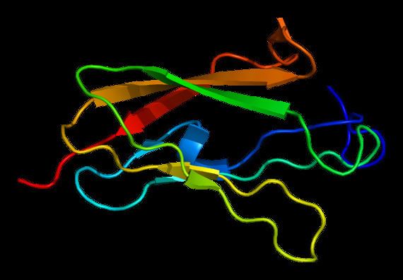
Structure
The protein structure includes an actin binding N terminal domain, 24 internal repeats and 2 hinge regions.
Interactions
Filamin has been shown to interact with:
RNA editing
The edited residue was previously recorded as a single nucleotide polymorphism(SNP) in dbSNP.
Type
A to I RNA editing is catalyzed by a family of adenosine deaminases acting on RNA (ADARs) that specifically recognize adenosines within double-stranded regions of pre-mRNAs and deaminate them to inosine.Inosines are recognised as guanosine by the cells translational machinery. There are three members of the ADAR family ADARs 1-3 with ADAR 1 and ADAR 2 being the only enzymatically active members.ADAR3 is thought to have a regulatory role in the brain. ADAR1 and ADAR 2 are widely expressed in tissues while ADAR 3 is restricted to the brain. The double stranded regions of RNA are formed by base-pairing between residues in a region complementary to the region of the editing site. This complementary region is usually found in a neighbouring intron but can also be located in an exonic sequence. The region that base pairs with the editing region is known as an Editing Complentary Sequence (ECS).
Site
The one editing site of FLNA pre-mRNA is located within amino acid 2341 of the final protein. The Glutamine (Q) codon is altered due to a site specific deamination of an adenosine at the editing site to an Arginine (R) codon. The editing region is predicted to form a double stranded region of 32 base pairs in length with a complementary sequence about 200 nucleotides downstream of the editing site. This ECS is found in an intronic sequence. Editing at the Q/R site is likely to involve both ADAR1 and ADAR2.Mice ADAR2 knockouts show a decrease in editing at the Q/R site.ADAR1 double knockouts have no effect on editing.
Structure
The edited adenosine is located in the 22 immunogloulin like repeat of the protein. This region is an integrin β binding domain and a RAC1 binding domain. The amino acid change is likely to effect the electrostatic potential of the binding domains. FLNA editing site is 2 nucleotides from a splice site like the R/G site of GluR-2. Both transcripts have 7/8 identical nucleotides around their editing sites. Since it is widely thought that editing at the GLUR-2 Q/R site influences splicing, the sequence and editing site similarity could mean that editing at the FLNA site could also regulate splicing. In vitro experiments of gluR-2 have shown that presence of ADAR2 results in inhibition of splicing. Analysis of EST data for FLNA show that there is a link between editing of the last exon codon and retention of the following intron.
Function
The change in electrostatic potential is likely to effect the binding of FLNA to the many proteins it interacts with.