
Nerve radial nerve | ||
 | ||
Artery posterior interosseous artery Antagonist Flexor digitorum superficialis muscle, Flexor digitorum profundus muscle | ||
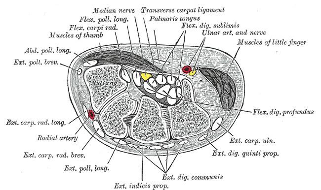
The extensor digitorum muscle (also known as extensor digitorum communis) is a muscle of the posterior forearm present in humans and other animals. It extends the medial four digits of the hand.
Contents
Structure
It arises from the lateral epicondyle of the humerus, by the common tendon; from the intermuscular septa between it and the adjacent muscles, and from the antebrachial fascia. It divides below into four tendons, which pass, together with that of the extensor indicis proprius, through a separate compartment of the dorsal carpal ligament, within a mucous sheath. The tendons then diverge on the back of the hand, and are inserted into the middle and distal phalanges of the fingers in the following manner.
Opposite the metacarpophalangeal articulation each tendon is bound by fasciculi to the collateral ligaments and serves as the dorsal ligament of this joint; after having crossed the joint, it spreads out into a broad aponeurosis, which covers the dorsal surface of the first phalanx and is reinforced, in this situation, by the tendons of the interossei and lumbricalis.
Opposite the first interphalangeal joint this aponeurosis divides into three slips; an intermediate and two collateral: the former is inserted into the base of the second phalanx; and the two collateral, which are continued onward along the sides of the second phalanx, unite by their contiguous margins, and are inserted into the dorsal surface of the last phalanx. As the tendons cross the interphalangeal joints, they furnish them with dorsal ligaments. The tendon to the index finger is accompanied by the extensor indicis proprius, which lies on its ulnar side. On the back of the hand, the tendons to the middle, ring, and little fingers are connected by two obliquely placed bands, one from the third tendon passing inferior and laterally to the second tendon, and the other passing from the same tendon inferior and medially to the fourth.
Occasionally the first tendon is connected to the second by a thin transverse band. Collectively, these are known as the sagittal bands; they serve to maintain the central alignment of the extensor tendons over the metacarpal head, thus increasing the available leverage. Injuries (such as by an external flexion force during active extension) may allow the tendon to dislocate into the intermetacarpal space; the extensor tendon then acts as a flexor and the finger may no longer be actively extended. This may be corrected surgically by using a slip of the extensor tendon to replace the damaged ligamentous band
Function
The extensor digitorum communis extends the phalanges, then the wrist, and finally the elbow. It tends to separate the fingers as it extends them.
In the fingers, the extensor digitorum acts principally on the proximal phalanges, acting to extend the metacarpophalangeal joint. Extension of the proximal and distal interphalangeal joints, however, is mediated predominantly by the dorsal and palmar interossei and lumbricals of the hand.