
Specialty infectious disease DiseasesDB 8472 MeSH D008271 | ICD-10 B47.0 Patient UK Eumycetoma | |
 | ||
eMedicine med/30 derm/280 derm/147 | ||
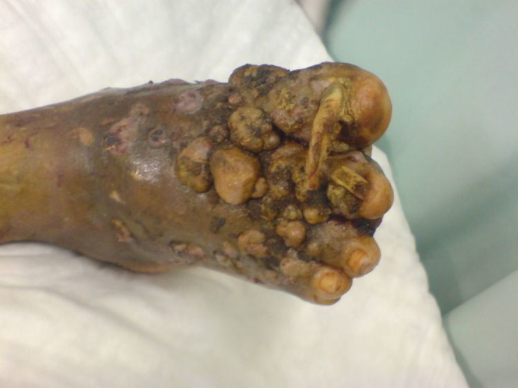
Eumycetoma is a chronic granulomatous fungal disease of humans, affecting mainly the limbs, and sometimes the abdominal and chest walls or the head. Mycetoma pedis (mycetoma of the foot), the most common form of mycetoma, is known widely as the Madura foot. The infection is endemic in Africa, India and the Central and South Americas.
Contents
Signs and symptoms
The initial lesion is a small subcutaneous swelling following minor trauma. Later, sinuses that discharge purulent and seropurulent exudates containing grains which are fungal colonies are formed. Destruction of deeper tissues, and deformity and loss of function in the affected limbs may occur in late stages.
Cause
Mycetoma may be caused by bacteria from the phylum Actinomycetes, or by fungi (Eumycetes) where it is called Eumycetoma. Bacterial and fungal species that can cause mycetoma are listed below under their characteristic colours of discharge from infected wounds:
Red discharge
White or Yellow discharge
Black discharge
Some species of the bacterial genus Nocardia (including Nocardia asteroides and Nocardia brasiliensis) which can cause mycetoma produce a yellow coloured discharge, and those of the bacterial genus Streptomyces (including Streptomyces somaliensis) produce an yellow or red coloured discharge.
Pathogenesis
The disease is usually seen in field workers like farmers, and generally affects men between 20 and 40 years. The disease is acquired by inoculation of grains of fungal spores from the soil through a breach in the skin produced by minor trauma like a thorn prick. The disease then spreads to deeper tissues and also forms sinus tracts leading to skin surface. Mature lesions are characterised by a grainy discharge from these sinuses. These discharges contain fungal colonies and are infective. Spread of infection internally through blood or lymph is uncommon.
Infections that produce a black discharge mainly spread subcutaneously. In the red and yellow varieties deep spread occurs early, infiltrating muscles and bones but sparing nerves and tendons, which are highly resistant to the invasion.
Botryomycosis, also known as bacterial pseudomycosis, produces a similar clinical picture and is caused usually by Staphylococcus aureus. Other bacteria may also cause botryomycosis.
Diagnosis
Diagnosis of mycetoma is usually established clinically in endemic areas. X rays and ultrasonography may be employed in evaluating the extent of the disease. X rays findings are extremely variable. The disease is most often observed at an advanced stage that exhibits extensive destruction of all bones of the foot. Rarely, a single lesion may be seen in the tibia where the picture is identical with chronic osteomyelitis. Cytology of fine needle aspirate or pus from the lesion, and tissue biopsy may be undertaken sometimes. Some publications have claimed a "dot in a circle sign" as a characteristic MRI feature for this condition (this feature has also been described on ultrasound).
Differential diagnosis
The following clinical conditions may be considered before diagnosing a patient with mycetoma:
- Tuberculous ulcer
- Kaposi's sarcoma, a vascular tumour of skin usually seen in AIDS.
- Leprosy
- Syphilis
- Malignant neoplasm
- Tropical ulcer
- Botryomycosis, a skin infection usually caused by the bacteria Staphylococcus aureus.
Prevention
No vaccine is available. Simple hygienic precautions like wearing shoes or sandals while working in fields, and washing hands and feet at regular intervals may help prevent the disease.
Treatment
Drugs like ketoconazole, voriconazole, and itraconazole are generally employed in treating the infection. Actinomycetes usually respond well to medical treatment, but the eumycetes are generally resistant and may require surgical interventions including amputation.
Epidemiology
The disease is endemic in tropical and subtropical regions. The exact incidence and geographical distribution of mycetoma throughout the world is not known as the disease is usually painless, slowly progressive and presented to health centres only in late stages by majority of patients. Mycetoma has an uneven worldwide distribution.
History
Madura foot or maduromycosis or maduramycosis, is described in ancient writings of India as Padavalmika, which, translated means Foot anthill. The first modern description of Madura foot was made in 1842 from Madurai (the city after which the disease was named Madura mycosis) in India, by Gill. The fungal etiology of the disease was established in 1860 by Carter.